Characterizing the Micro-Mechanical Properties of Soft Materials, Tissues and Cells

Complete the form below to unlock access to ALL audio articles.
This special promotional feature was prepared by Optics11.
The field of tissue engineering and regenerative medicine (TERM) has expanded rapidly the last decade, driven by the aim to improve quality of life by advancing healthcare. The process of generating functional human tissue from cells, in a laboratory or clinical environment, has many important factors, such as the biochemical composition of the cellular environment. However, one important factor that has been recognized more recently to play an important role in cell differentiation and tissue formation is the mechanical properties of (bio)material grafts, substitutes and scaffolds.
Traditional trade-off
Characterizing the mechanical properties of soft (bio)materials is traditionally a trade-off between measuring the bulk mechanical properties, i.e. by rheology, bulk compression or tensile testing, and methods that enable investigating local mechanical properties, i.e. traditional (micro-, nano-) indentation or atomic force microscopy (AFM). In tissue engineering and regenerative medicine this trade-off is even more stressed, as the need for local interrogation is combined with the need for high sensitivity, compatibility and ease of use with biological samples, e.g. measuring in liquid environments. Where AFM’s typically match high sensitivity, they can be complex to operate and setting up an experiment can be time consuming due to extensive alignment and calibration procedures.

Combining the best of two worlds
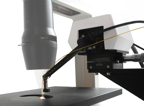
To enable mechanical studies for tissue, cell and biomaterial researchers, Optics11 has designed the Piuma Nanoindenter, which captures the benefits of both traditional nanoindenters and AFM solutions, but not their limitations. The Piuma Nanoindenter uses a novel proprietary optical interferometric technology in combination with a monolithical cantilever probe that enables the automated characterization of the local mechanical properties of the softest materials with high sensitivity, speed and ease of use.
"The Piuma opened a new world for us. We now are able to perform measurements on very low stiffness tissues, with a precision that allows us to study embryos. Colleagues are impressed and come with other challenges that the Piuma easily meets." – Prof. dr. ir. Theo Smit, Dept. Orthopedic surgery, VU University Medical Centre, Amsterdam, The Netherlands.

Many applications
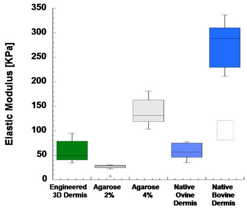
The Piuma is currently being used in a number of key universities and research institutes, improving graft and scaffold characteristics and contributing to the understanding of mechanical processes down to the cell level. At present, the Piuma enables researchers to advance the engineering and regeneration of dermis, cartilage, cornea, bone and many other tissues, as well as (stem)cell development in environments with specific mechanical properties. Typical scaffold materials such as hydrogels and polymers are characterized routinely. In addition, the Piuma is used for studying disease models, such as brain conditions, liver conditions, eye conditions, and many more. In general the Piuma is contributing to an improved quality of life by advancing researchers’ understanding of the mechanical properties of (bio)materials, tissues and (stem)cells.
"The Piuma is a very powerful, reliable, and use-friendly instrument to assess the mechanical properties of both living tissue (e.g. tissue explants) and in vitro engineered tissue." Francesco Urciuolo, PhD - Research Technologist at Instituto Italiano Tecnologia, Naples, Italy.

Maximal flexibility
Sample mounting is made easy with the Piuma system: it fits almost any container, petri dish or multi-well plate up to 12 wells. If you require more flexibility, for example when combining nanoindentation with other analysis instruments, such as optical microscopes, this is also possible with the Piuma Chiaro. This enables not only the mechanical characterization of single cells, as well as investigating specific or very local sites of tissues and scaffolds.

Author Biography Ernst Breel, MSc (27), Applications Specialist at Optics11, helps researchers to set up new experiments to improve their understanding of the optical and mechanical properties of biomaterials, tissues and cells. He recently graduated from the VU University in Amsterdam with a masters’ degree in Physics, track Science, Business & Innovation.
Request more information
Currently Optics11 provides on-site demonstrations of the Piuma Nanoindenter in various countries. In case you would be interested in a demonstration, or would like to request more information about the Piuma Nanoindenter, please contact Ernst Breel.


