DETERMINATION OF HOMOCYSTEINE THIOLACTONE IN URINE BY FIELD AMPLIFIED SAMPLE INJECTION AND SWEEPING MEKC METHOD WITH UV DETECTION
Homocysteine-thiolactone (HTL) is a cyclic thioester of homocysteine, which is synthesized by methionyl-tRNA synthetase, that mistakenly activates cyclization of homocysteine. HTL is highly reactive and readily reacts with ε-amino groups of protein lysine residues in a process called N-homocysteinylation. This reaction alters the proteins structure and function and contributes to the emergence of many diseases. Because of its low concentration in biological fluids it is important to choose the determination technique outstanding high sensitivity and low operating cost.
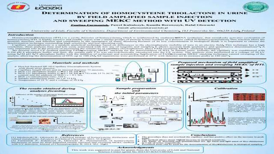
Capillary electrophoresis is a modern analytical technique based on differences in the electrophoretic mobility of ions in an electric field. This technique has a high resolution, short analysis time and low costs associated with low use of reagents and samples. It allows accurate both qualitative and quantitative determinations[1]. A very important role in the process of increasing the sensitivity in capillary electrophoresis, plays FASI-sweeping MEKC stacking technique.
We present novel CE method utilizing FASI-sweeping MEKC stacking for the determination of HTL in human urine. Initially, the capillary was conditioned with 0.1M pH 6 phosphate buffer and then, the sample (pH 5.5) was electrokinetically introduced into the CE system. The analysis were performed at a voltage of 20 kV using 0.1M pH 4.75 phosphate buffer with the addition of surfactant and acetonitrile. Application of FASI-sweeping MEKC stacking technique caused sweeping of the analyte, allowing the determination of HTL up to 100 nM. All separations were performed at 25 C with detector set to measure peaks at 240 nm[2].


