Nexcelom Bioscience Launches 5 Channel Celigo Image Cytometer
Complete the form below to unlock access to ALL audio articles.
Nexcelom Bioscience has announced that it has released the addition of a 5th channel available as an option on the Celigo image cytometer. The Celigo is a bench-top image cytometry system providing whole-well imaging and quantitative data, through image analysis for demanding cell-based analytical applications. Previous configurations allowed bright field only imaging, or bright field imaging plus 3 fluorescent channels. This new option of bright field plus 4 fluorescent (red, green, blue and far-red) channels will allow researchers more capabilities without having to compromise their assays.
This addition to the product offering was driven by the requests of researchers and current customers. Researchers now use the Celigo image cytometer to develop cell-based assays with cells expressing GFP and RFP in combination with blue and far-red dyes. The other configuration options will still be available for order – allowing researchers to customize the Celigo to best meet their needs.
“The addition of a 5th fluorescent channel on the Celigo responds to the increasingly wider array of fluorescent reagents for cell-based assays available to researchers today and provides our customers with additional multiplexing capabilities for the acquisition and analysis of more complex cell populations.” said Olivier Déry, Director of Celigo Business at Nexcelom Bioscience.
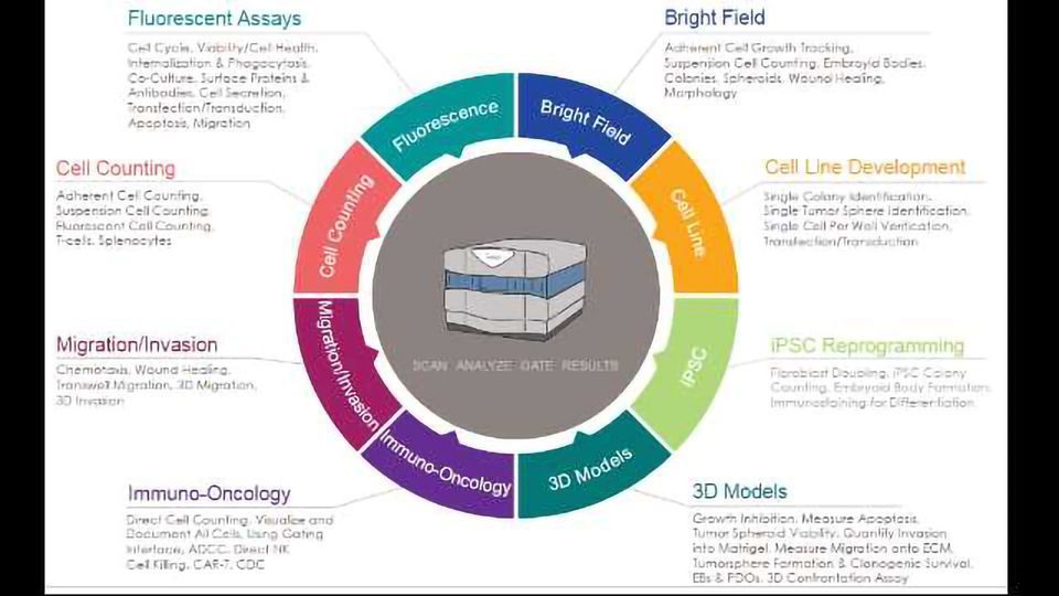
The Celigo allows users to perform high-speed, fully automated imaging and quantification of a wide range of cell types across complex sample types. It enables an extensive menu of applications, and with the option to have it configured with bright field and four fluorescent channels will open up greater possibilities for its use and applications within fields such as immuno-oncology, drug discovery and many others.

