Cross-linking Mass Spectrometry: A Key Player in the Structural Biologist's Toolbox

Complete the form below to unlock access to ALL audio articles.
Single proteins, protein complexes, and the interactions between proteins are a fundamental component of the cellular processes that underlie the physiological function of an organism. Structural biology places emphasis on blueprints; if we understand and can deconstruct the blueprint of a molecule or complex, then when that molecule malfunctions, we can repair it.
Protein structure information has been collected by scientists almost exclusively using methods such as X-ray crystallography and nuclear magnetic resonance (NMR) spectroscopy – as of 2018, 89% of protein data bank entries were contributed by studies using x-ray crystallography and 9% using NMR.1
Unsurprisingly, as the proteomics field has grown over recent years, in parallel, so has the size and complexity of the molecules that scientists want to study.2 The latest addition to the structural biologist's toolbox, cross-linking mass spectrometry (XL-MS), is helping researchers achieve these goals: "In general: “difficult” protein complexes can be studied where NMR or X-ray fail," says Andrea Sinz, Professor in the Department of Pharmaceutical Chemistry and Bioanalytics at The Charles Tanford Protein Center (CTP). Here, we take a brief look at the basic principles of XL-MS before speaking to experts in the field about how it is being applied.
How does XL-MS work?
"In XL-MS, we use chemical reagents and/or UV light to form covalent bonds between side chains of amino acids," explains Alexander Leitner, PhD, Senior Scientist for the Aebersold Group at the Institute of Molecular Systems Biology.
At the basic level, proteins consist of amino acid chains linked together by peptide bonds. Non-covalent bonds, such as electrostatic and hydrophobic interactions and van der Waals forces also contribute to the various levels of protein structure. Note: these non-covalent bonds are difficult to detect, hence the requirement for chemical reagents in XL-MS.
"The main differences (in addition to the cross-linking step) between MS and XL-MS are in MS data acquisition and data analysis, because we are always looking at two peptides connected to each other by the cross-link," says Leitner. "The cross-links tell us that two reactive sites are close to each other in space, either on the same protein or on different ones. This way, we can learn about the three-dimensional arrangements of proteins and protein complexes, and even about larger protein networks."
Due to recent technological advances, an array of workflows exists for XL-MS experiments, varying the cross-linking reagents and bioinformatic tools utilized as outlined by Yu and Huang in their comprehensive 2018 review.1
Thus far, XL-MS has characterized the structure of several large assemblies such as the proteasome, RNA polymerase and chromatin remodeling complexes.3 "Overall, I see three main application areas of XL-MS", comments Leitner. "Single proteins, where XL data is helping to determine the fold," he continues, "Protein complexes, where XL is used alone or in combination with other biophysical and structural biology methods to study the architecture of the assemblies, and entire protein networks in whole cells or cell lysates, where one can obtain insights about interactions and their changes at the systems level." Let's explore these applications further.
Integrative structural methods
The field has recently witnessed researchers opting to marry XL-MS with cryogenic electron microscopy (cryo-EM). The result? A structural biology power couple.

The study findings showed a structural and functional basis for TRiC-PFD cooperation in protein folding. An electrostatic interaction allows PFD to bind to the open TRiC state and pivot to a conformation that aligns their two substrate-binding chambers, altering the chemical properties of the substrate environment. This physical contact between TRiC and PFD is essential for effective proteostasis and disrupting such contact leads to PFD being rendered toxic and the accumulation of misfolded amyloid aggregates. Of the research Leitner comments: "This is a very good example for how XL-MS can interface with cryo-EM and biochemical/functional assays to understand how large complexes work, in this case to fold their substrates."
The focus of Sinz's laboratory at the CTP is the development of novel MS-cleavable cross-linkers.
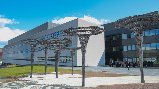
The Charles Tanford Protein Center of Martin Luther University Halle-Wittenberg. The center is dedicated to protein research. Image credit: Andrea Sinz.
Sinz and colleagues recently published their proteome wide XL-MS workflow using the MS-cleavable crosslinker, disuccinimidyl dibutyric urea (DBSU).5,6 DBSU contains a urea linker that can be cleaved during the gas phase of tandem MS via collision-induced dissociation (CID), allowing MS3 acquisition. Accompanying the novel workflow, the team upgraded the freeware tool MeroX 2.0 so that it can be used to conduct fully automated and reliable large-scale protein-protein XL analyses. "Using MS-cleavable cross-linkers is really beautiful as the cleavage gives you a signature, and the signature is crucial to study the cross-links in vivo or in whole organisms," remarks Sinz.
The researchers applied their workflow to capture protein-protein interactions (PPIs) at a system-wide level in Drosophila embryos undergoing maternal-to-zygotic transition. The Drosophila embryo at age 0-2 h is transcriptionally silent, relying on maternal RNA and proteins provided in oogenesis. This means that any processes occurring in the embryo prior to transcriptional genome activation are regulated at the post-transcriptional level:
The results of XL-MS analyses using MS-cleavable cross-linkers revealed 1611 interprotein cross-linking sites and 5825 intraprotein cross-links that defined the topology of novel protein complexes in early Drosophila embryogenesis. "PPIs identified in this system might aid the understanding of the complex nature of regulation during early embryonic development," the researchers note.
Challenges in a maturing field
As discussed, XL-MS has played a significant role in recent integrated structural biology research. It has major advantages compared with other techniques in that it tolerates a wide range of buffer conditions, allows for sample heterogeneity and requires samples in small quantities.7 Nonetheless, there is always room for improvement says Sinz: "The main challenge is in software development. We need to develop software tools that can handle the complicated peptides and cross-linking product mixtures. Software development has made huge progress. Currently the challenge is to have novel reagents and cross-linkers that are cleavable in the mass spectrometer to get the signatures for automated analysis."
Of course, the addition of novel analysis tools adds complexity to an already highly technical method, as acknowledged by Leitner: "One thing that may be intimidating to newcomers to the field is the huge diversity of the cross-linking chemistries, experimental protocols and software tools." He adds, "Almost all specialized labs initially developed their own protocols and software pipelines. One reason for that is the versatility of the method and the diversity of the applications. This makes it difficult for inexperienced users to make proper and objective comparisons and figure out what the best strategy for their particular use case would be."
A look to the future of XL-MS
Sinz believes that there are two routes at the horizon of XL-MS:
- Application with cryo-EM for solving partial, complementary structures to analyze very large protein assemblies or membrane protein complexes
- Mapping protein interaction networks on the system-wide level
Her thoughts are echoed by Leitner: "I think it is unlikely that a one-size-fits-all solution will emerge in terms of chemistry or experimental workflow. Progress in all three main focal areas (proteins, complexes, proteomes) will certainly continue, as will the integration of XL-MS with data from other sources."
As for the complex nature of XL-MS, the researchers are optimistic that the structural insights achieved through XL-MS studies will soon be obtainable for a wider-range of scientists: "Slowly, the community is getting together to carry out benchmark studies and to define reporting standards and data formats. This will increase transparency for everyone; it is just a process that takes time," Leitner concludes.


