Cell Migration: Clinical Relevance, Unique Movement Patterns and Driving Technologies

Complete the form below to unlock access to ALL audio articles.
Cells like to move it, move it! Understanding the mechanisms behind cell migration represents an important piece to the puzzle of many fundamental biological processes and pathologies. This article delves into textbook models of cell migration, recent insights on movement patterns and governing mechanisms, and highlights the technology driving cell migration research forward.
Cell migration and its importance in health and disease
Cell migration is a broad term used to describe the translation of cells from one location to another. There are many fundamental biological processes that rely on this, such as embryonic development1, wound healing2, and fertilization.
On the flipside, the migration of cells also plays a major role in pathological development, such as cancer metastasis3 and autoimmune disorders. Interest in understanding cell migration has been boosted by the rise in regenerative medicine, and is predicated on the hope that knowledge of the precise mechanisms underlying cell migration can be leveraged to create novel therapeutic approaches.
Brendon Baker, assistant professor of biomedical engineering at the University of Michigan notes that there are many regenerative medicine approaches that could be aided by improving our understanding of the cellular microenvironment and its influence on cell migration. He lists a few examples:
“Stem cell-based therapies require mobilizing and targeting cells to intended sites of repair, whether the goal is integration of differentiated stem cells with damaged tissues or delivery of cytokines and growth factors to induce a localized regenerative response.
Being able to control cell migration would also be relevant to the design of dermal wound healing grafts for burn victims or diabetic patients, where ideally cells are guided from nearby healthy tissues to invade into the graft to orchestrate a complex series of cell-type specific processes – including the formation of new vasculature by endothelial cells, production or restoration of the matrix by fibroblasts, and re-epithelialization by keratinocytes.”
Cell migration research has benefited greatly from leaps in imaging techniques and bioengineering, revealing fascinating insights into the way cells move, and the mechanisms that drive their motility.
What the textbooks told us about cell migration
While prokaryotic organisms and sperms cells can use flagella or cilia to propel themselves forward, eukaryotic cell migration generally relies on drastic changes in cell shape which are driven by the cytoskeleton. One of the major migratory behaviors displayed by cells is known as the textbook model of mesenchymal cell migration.4
In mesenchymal migration, motile cells exhibit a leading edge of the cell, which is thought to be driven by the extension of a branched network of actin filaments that polymerize beneath the plasma membrane.5 The leading edge extends, and adhesion points enable the cell to secure contact with its surface. Simultaneously, adhesions at the rear of the cell must disassemble to enable productive forward movement of the cell.
There are two main models of cell motility that have been put forward over the decades, which largely differ in the way that the trailing edge of the cell disassembles and reassembles during the retraction phase of a cell’s motion. The models are described here, and in the video below:
- Cytoskeletal model of movement: Actin proteins polymerize quickly to form filaments and branched networks at the leading edge of the cell, which pushes the cell membrane forward. Simultaneously, microtubules at the trailing edge of the cell carry out two main functions which enable the cell to be dynamic; by acting as a rudder to steer the cell, and by acting as an anchor to stop the cell from moving.
- Membrane flow model: In this model, sections of the plasma membrane endocytose from the back of the cell, and move to the front of the cell. Some vesicles are made solely of plasma membrane, and others contain integrin proteins which attach the cell cytoskeleton to the extracellular matrix.
Video credit: Khan Academy
Whether both models come into play, or only one, is still a matter of dispute.4
Amoeboid movement is another major category of cell migration, which refers to the movement of rounded or ellipsoid cells that lack mature focal adhesions – the proteins that form mechanical links between intracellular actin and the extracellular matrix.6 There are two subtypes of amoeboid movement, which are defined by whether cells pull on substrate (usually a weak interaction) or not (a propulsive, pushing mode of action). Amoeboid movement came into the spotlight recently, when dendritic cells were shown to probe different pathways before picking the widest path, when navigating route options in a microfluidic model.7 The positioning of the nucleus was found to be fundamental to the strategy of locomotion – by positioning the nucleus at the front, cells could used their bulkiest compartment as a mechanical gauge. Smaller cytoplasmic protrusions were shown to retract from smaller pores, once the nucleus and closely associated microtubule organizing center had passed the largest pore.
Watch: How White Blood Cells Choose the Path of Least Resistance
Unconventional movement patterns revealed
Who would have thought cells could propel themselves forward like a slingshot?!In stark contrast to mesenchymal cell migration, human fibroblast cells have been observed applying forces to stretch the surrounding matrix, and subsequently “slingshot” themselves forward.8 This distinct movement results in cells traveling more than five times faster than previously documented.
The observation came from a research group from the University of Michigan, who developed 3D matrices to mimic the fibrous architecture characteristics of native tissues. The alignment and stiffness of these biomaterials could be tuned, enabling the group to explore how the properties of stromal tissue could hinder or promote cell migration.
In stiff matrices, cells movement was consistent with the mesenchymal model of cell migration. However, cells in deformable matrices showed a different approach, and stretched the matrix fibers to store elastic energy. Consequently, the eventual loss of adhesion at the rear of the cell induced sudden matrix recoil and with it, rapid cell translocation.
The extent to which the matrices compare to those found in vivo is difficult to assess, but Baker’s lab group tuned the mechanics over a range to match values of tissues reported by groups in the literature.
Video credit: Baker lab, University of Michigan
This work highlights the possibility of designing biomaterials that rapidly recruit repair cells to wound sites, at a rate that could aid the transplant success of tissues. To achieve this, more than matrix stiffness will need to be optimized. Baker elaborates on other considerations:
“For regenerative therapies, we need to think about much thicker, more three-dimensional structures and this will require considering pore size. In my graduate work, we showed that thick scaffolds composed of similar fibers generally prevent cells from invading into and colonizing the material unless sufficient pore size is engineered into the material.
Another feature we touch on in this work is contact guidance – which is how structural cues guide directed cell migration (migration in a prescribed direction). Thinking about large 3D scaffolds for regenerative medicine applications, we would need to engineer in guidance cues that support invasion into the material. So not only should we consider the mechanics of biomaterials, but structural cues that direct cell migration and sufficient porosity to afford cells the ability to move, with a key challenge being that these three aspects are co-dependent.”
Master proteins coordinating structural changes
Understanding the structural cues that direct cell migration has been a focus for Daniel Lietha, a principal investigator who recently moved his group to the Biological Research Center (CIB) of the Spanish Research Council (CSIC) in Madrid, Spain. In previous work at the Spanish National Cancer Research Center (CNIO), his group was focused on understanding the changes that prompt conformational changes in focal adhesion kinase (FAK), a key signaling molecule regulating cell adhesion, migration and survival.FAK drives signals that are essential for focal adhesion formation and the formation of lamellipodia – the “foot” present at the leading edge of the cell. When inactive, FAK is diffused in the cytoplasm, and can be found activated at focal adhesions.
Lietha explains the rationale behind establishing exactly what stimulates FAK activation:
“If you know how FAK is activated, you might be able to prevent this from happening in invasive tumors, where FAK signaling is key. It is also interesting from a scientific point of view. It has been long known that force applied by actomyosin contraction activates signals in focal adhesions. But how the mechanical force is converted in a biochemical signal and what constitutes the force sensor, was unknown.”
Hypothesizing that FAK can act as a sensor of changes in the extracellular and intracellular framework, Lietha and his team used atomic force microscopy (AFM) to simulate the forces generated by the contraction of actin filaments in the cytoskeleton.9 By combining their measurements with structural information of FAK and force-probe molecular dynamics simulations, the unfolding of defined structural features in FAK could be explained by measured changes in force. Lietha explains how AFM was utilized in this study:
“Single molecules (FAK in our case) can be attached between the AFM surface and cantilever, and extended with nanometer accuracy – while force is measured with piconewton accuracy.”
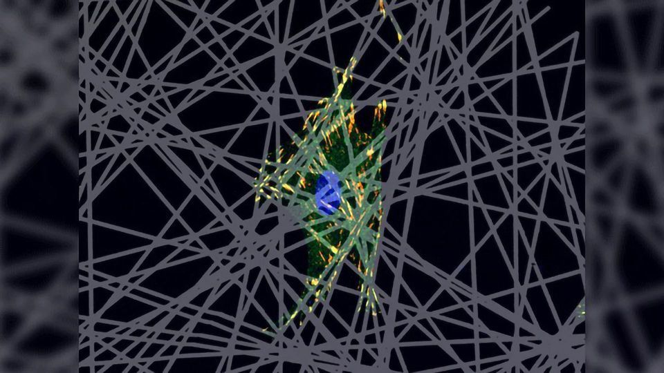
 Left: During cell adhesion to the extracellular matrix (ECM) force is generated in the focal adhesion complex. FAK is exposed to force through docking of its N-terminal FERM domain to the plasma membrane and its C-terminal FAT domain to cytoskeletal focal adhesion proteins. Middle: Stretching forces on FAK were mimicked in an atomic force microscopy (AFM) setup by attaching FAK molecules via tags to the AFM cantilever and surface.
 As FAK is frequently overexpressed in cancer, these findings are particularly relevant for the design of anticancer drugs that inhibit the catalytic and scaffolding functions of FAK.
Diverse research approaches are propelling cell migration research forward
Like many other areas of biology, cell migration research is benefiting greatly from technical advances across disciplines. While gene-editing tools such as CRISPR allow studies of specific roles of genes in metastasis10, simplified models remain relevant. The fish epidermal keratocyte, wound-healing cells in fish skin, have proven to be valuable for research – partly because their simple shape allows the description of basic principles that govern their movement.11 As fish epidermal keratocytes move rapidly, great insights can be gained through single particle tracking of diffusing particles.12Advances in 3D imaging have allowed these simplified models to be revisited; in 2018, Okimura et al. described the specific arrangement of fibers key to migration velocity of crawling keratocytes.13 The development of fluorescent probes allow fluorescently labeled cells to be tracked in real-time, either in explant cultures or directly intravitally in live animals.14
Artificial environments such as tunable engineered matrices and microfluidic platforms have enabled researchers to ask specific questions about how cells navigate their way through tissues, and imaging advances have revolutionized our ability to find the answers.
References:
- Reig, G., Pulgar, E., Concha, M. L. (2014). Cell migration: from tissue culture to embryos. Development 141: 1999–2013
- Li, L., He, Y., Zhao, M., Jiang, J. (2013). Collective cell migration: implications for wound healing and cancer invasion. Burns Trauma 1(1):21–26
- Bravo-Cordero, J. J., Hodgson, L., Condeelis, J. (2011). Directed cell invasion and migration during metastasis. Current Opinion in Cell Biology 24(2): 277–283
- Innocenti, M. (2018). New insights into the formation and the function of lamellipodia and ruffles in mesenchymal cell migration. Cell Adhesion & Migration 12(5): 401–416
- Pollard, T., Borisy, G. (2003). Cellular motility driven by assembly and disassembly of actin filaments. Cell 112(4): 453–465
- Friedl, P., Wolf, K. (2009). Plasticity of cell migration: a multiscale tuning model. The Journal of Cell Biology 188(1): 11–19
- Renkawitz, J., et al. Nuclear positioning facilitates amoeboid migration along the path of least resistance. Nature 568: 546–550
- Wang, W., Davidson, C., Baker, B. (2019). Actomyosin contractility-dependent matrix stretch and recoil induces rapid cell migration. Nature Communications 10: 1186
- Bauer, M., et al. (2019). Structural and mechanistic insights into mechanoactivation of focal adhesion kinase. Proceedings of the National Academy of Sciences of the United States of America 116(14): 6766–6774
- Van Treuren, T., Vishwanatha, J. (2018). CRISPR deletion of MIEN1 in breast cancer cells. PLOS One 13(10):e0204976
- Lee, J., Ishihara, A., Jacobson, K. (1993). The fish epidermal keratocyte as a model system for the study of cell locomotion. Symp Soc Exp Biol 47:73–89 https://europepmc.org/abstract/med/8165580
- Kucik, D. F., Elson, E. L., Sheetz, M. P. (1990). Cell migration does not produce membrane flow. The Journal of Cell Biology 111: 1617-1622 http://jcb.rupress.org/content/jcb/111/4/1617.full.pdf
- Okimura, C., Taniguchi, A., Nonaka, S., Iwadate, Y. (2018). Rotation of stress fibers as a single wheel in migrating fish keratocytes. Scientific Reports 8: 10615
- Benechet, A., Menon, M., Khanna, K. (2014). Visualizing T cell migration in situ. Frontiers in Immunology 5:363

