Mycoplasma Contamination: Minimizing Risk To Avoid Cell Culture Complications
Credit: Thermo Fisher Scientific
Want to listen to this article for FREE?
Complete the form below to unlock access to ALL audio articles.
Read time: 6 minutes
Avoiding contamination is a serious and continual challenge across all cell culture processes. In a previous article, we examined ways of minimizing the impact of chemical contamination resulting from extractables in plasticware. This follow-on piece explores a problematic microbiological contaminant, mycoplasma, and examines the tools available to help prevent its establishment and mitigate its impact.
Microbial contamination of mammalian cell culture not only threatens the viability of the culture itself, it raises questions about the integrity of the process and validity of the resulting data or products. The nature of the cell culture environment means it coincidentally satisfies the nutritional and growth requirements of numerous different microorganisms, which can all pose a potential risk. As a result, strict aseptic techniques must be applied alongside the implementation of a range of measures designed to help prevent or quickly detect and eliminate any contamination.
Among the many potential contaminants, mycoplasma species present a particular challenge. This is principally because their extremely small size makes them both difficult to exclude from the cell culture environment and hard to detect. Understanding the risks involved helps guide mitigation strategies, which include rigorous asepsis, intensive filtration of reagents and regular use of the most effective detection methods.
The problem with mycoplasma
Mycoplasmas are the smallest known free-living organisms. They have long been recognized as common contaminants of mammalian cell cultures, and remain a significant challenge in today’s laboratories and biopharmaceutical production facilities. In a 2015 study designed to obtain an unbiased assessment from multiple laboratories, analysis of sequence data from 9,395 rodent and primate samples from 884 series in the NCBI Sequence Read Archive, showed 11% were contaminated with mycoplasma.1
Mycoplasmas are distinguished from other bacteria by their extremely small physical size (0.15 - 0.3 µm), small genome (580 to 2,200 kb) and lack of a cell wall. The combination of their size and pleomorphism enables these organisms to pass through standard 0.2 µm sterilizing grade filters. Since they can then reach high concentrations without creating noticeable turbidity, the effects of contamination are not usually visible to the naked eye until the damage is done. The absence of a cell wall means they are not susceptible to commonly used cell culture antibiotics, making eradication difficult. To complicate matters further, antibiotics that are effective may be toxic to the cell culture itself.
Contamination with mycoplasma can have several different origins. Laboratory personnel are seen as a significant source with more than half of all mycoplasma infections in cell cultures resulting from organisms typically found in the human oropharyngeal tract.2 Animal-derived cell culture additives, and especially plant-based materials, can all give rise to other types of mycoplasmas. Existing contaminated cell lines and equipment also pose a serious threat.
Set the Standard for Quality and Superior Performance While Protecting Valuable Research

Mycoplasma contamination is a significant challenge in today's laboratories. Undoubtedly, prevention is better than remediation, and high filtration rate and high throughput filter systems make it easier for laboratories to implement preventative strategies. Our products are designed to effectively support your entire filtration process and ensure that you achieve the most accurate results.
Discover MoreSponsored Content
Mycoplasma contamination may persist undetected for many years in cell cultures.3 While it does not always cause cell death, it can alter the behavior of host cells. This may result in changes to cell metabolism and morphology, chromosomal damage and aberrations, and an increasing tendency to undergo apoptosis. There may be decreased rates of cell proliferation, reduced saturation density and agglutination in suspension cultures.
As a result, it is not possible to trust the experimental data from contaminated cultures, making re-work an absolute requirement. Once a laboratory has a confirmed mycoplasma contamination it will also need to undertake deep disinfection of all labware, equipment and surfaces. Often, large amounts of material that may have become contaminated through contact must be discarded. Mycoplasma contamination of cell cultures, therefore, has enormous financial and productivity implications for laboratories and production facilities.
Preventative strategies
Managing the risk of mycoplasma contamination relies on sustaining the highest levels of aseptic technique and good hygiene practice. Taking one or more of the following measures is also recommended: filtering all cell culture materials to below 0.1 µm, rather than the more conventional 0.2 µm, which misses mycoplasma; purchasing only 0.1 µm pre-filtered materials; regular testing with mycoplasma detection techniques that enable early action when contamination occurs.
Historically, filtration of cell culture media and components has been carried out using filters with a pore size of 0.2 µm, but there is now a shift towards 0.1 µm. A key driver behind these tighter specifications is the increasing move towards plant-based cell culture materials, which are in fact more prone to mycoplasma contamination than those derived from, for example, bovine serum albumen (BSA).4 Image 1 shows a typical vacuum filter set-up in which a modern design ensures the high filtration efficiency, fast flow rates and high throughput that are essential to maintaining productivity in cell culture laboratories, while playing a key role in mitigating contamination risks.
 Image 1: A typical filtration setup applicable to cell culture materials using a pore size of 0.1 µm. Credit: Thermo Fisher Scientific.
Today, an expanding array of media and cell culture components are offered as pre-filtered products. When purchasing these it is important to ensure all materials have been filtered to 0.1 µm. Not unexpectedly, such products can command premium prices. Those facilities using less-common cell culture media, custom media or media with additional components cannot always obtain pre-filtered products and so must undertake filtration on-site. Many people opt for a combination of approaches.
The importance of regular testing
Taking preventative measures to avoid mycoplasma contamination offers the best practice in managing cell cultures, and regular testing to check the effectiveness of these measures is crucial. The only assured way of detecting mycoplasma contamination is by testing cell cultures directly.
All testing is essentially reactive; once mycoplasma contamination is identified, the culture is already ruined and any results must be discarded. This problem is exacerbated by the challenges of using traditional microbiological tests for mycoplasma. These are lengthy and results may take up to 28 days. The latest rapid methods, however, support swifter confirmation of contamination so researchers know immediately to discard a cell culture batch, take the necessary remedial disinfection steps and start again.
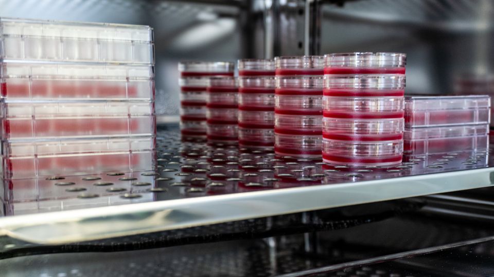
Such damage limitation means less work is done that subsequently needs repeating. PCR-based DNA testing is now an accepted method for detecting mycoplasma in cell culture and provides comprehensive results in just a few hours. Image 2 shows typical sampling points throughout a cell culture process. This early detection not only minimizes the re-work needed, but also allows rapid action to protect against the spread of mycoplasma into downstream equipment, processes and media.
 Image 2: Sampling points for mycoplasma. Rapid PCR-based testing for mycoplasma infection can be conducted throughout the cell culture manufacturing process, from inoculation through harvest. Credit: Thermo Fisher Scientific.
In conclusion
Mycoplasma contamination is an insidious problem in mammalian cell culture. Risk mitigation requires the adoption of strict aseptic practice, and is supported by the application of stringent media filtration criteria and the routine use of PCR-based rapid testing.
Undoubtedly, prevention is better than remediation. More effective filtration of media and components down to a membrane pore size of 0.1 µm is becoming the norm, avoiding problems associated with using standard 0.2 µm sterilization grade filters, which fail to prevent the passage of mycoplasma. High filtration rate and high throughput filter systems make it easier for laboratories to implement preventative strategies.
Alongside this is the growing commercial availability of pre-filtered materials. These validated and certified materials remove the need for further filtration. For many though, pre-filtered cell culture media may not be an option. On-site filtration is still required for custom media and specialist materials, for example, or to meet local specifications.
Finally, the advent and acceptance of rapid tests for mycoplasma means that laboratories can easily conduct regular testing of cell culture and know almost immediately if there is a contamination issue. They can then act fast to remove affected cultures and prevent further spread. Rapid testing – especially compared with the traditional 28 days to results with standard microbiological tests –minimizes lost work and the need for re-work.
References:
- Olarerin-George AO, Hogenesch JB. Assessing the prevalence of mycoplasma contamination in cell culture via a survey of NCBI's RNA-seq archive. Nucleic Acids Res. 2015;43(5):2535-2542. doi: 10.1093/nar/gkv136
- Nikfarjam L, Farzaneh P. Prevention and detection of mycoplasma contamination in cell culture. Cell J. 2021;13(4):203-212. PMCID: PMC3584481
- Uphoff CC, Drexler HG. Detecting mycoplasma contamination in cell cultures by polymerase chain reaction. In: Cree I. (eds) Cancer Cell Culture. Methods in Molecular Biology (Methods and Protocols). 2011. Humana Press. doi: 10.1007/978-1-61779-080-5_8
- Akers J, Meltzer T, Jornitz MW. The reoccurrence of mycoplasma contamination: Prevention strategies. Bioprocess International. https://bioprocessintl.com/downstream-processing/filtration/the-reoccurrence-of-mycoplasma-contamination-prevention-strategies-184084/. Published February 1, 2009. Accessed November 17, 2021

