Building an Atlas of the Brain To Guide Neuroscience: An Interview With the Allen Institute's Ed Lein

Complete the form below to unlock access to ALL audio articles.
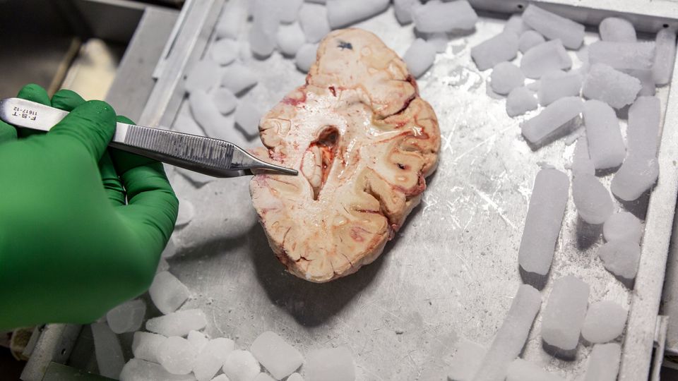
A new project to map the billions of cells in the human brain is underway.
The project, part of the National Institutes of Health’s Brain Research Through Advancing Innovative Neurotechnologies (BRAIN) initiative, involves 18 institutions and will be spearheaded by a team based at the Allen Institute for Brain Science in Seattle, WA, a subdivision of the Allen Institute.
Ed Lein, a senior investigator at the Institute, will co-lead the project alongside Hongkui Zeng, PhD, the Institute’s executive vice president and director.
The project, dubbed BICAN (BRAIN Initiative Cell Atlas Network) will exploit rapid advances in genomic and cellular technology to build an atlas that Lein hopes will provide a blueprint for decades of future brain research.
Technology Networks spoke to Lein to find out more about BICAN and how it could solve some of the biggest mysteries that surround our incredibly complex brain.
Ruairi J Mackenzie (RM): This isn’t the Allen Institute’s first brain atlas – how is BICAN different?
Ed Lein (EL): Our atlases have taken many forms over the years. They're mostly molecular atlases. We started with a mouse brain atlas, where we mapped the expression of all genes in the genome across the mouse brain. My interests have moved, over time, to study the human brain, and to try to bring to bear new technologies that can handle the scale of the fine structure and function of the human brain. This project that we're talking about is the latest and greatest iteration of this attempt to make a human brain atlas.
RM: Can you give an overview of the project?
EL: The philosophy that we took with this project is that this is a grand challenge to try to map the brain. And we want to not only create this map but make it in a way that's maximally useful for the neuroscience and medical communities.
Our atlas is just one of the projects within this bigger program. The concept is that we are now able to understand the cellular makeup of very complex tissues using the tools of genomics – in particular, a technique called single-cell or single-nucleus genomics. It lets you measure all the genes that are being used in individual cells. This can be scaled up dramatically; you can do this now across many millions of cells, each with a unique genetic signature. These genes give the distinct kinds of cells their specific properties. It turns out that if you can measure all the genes in a cell, you can categorize those cells on the basis of the genes that they use. You can make a classification and take this really complex tissue and understand all of the very specific kinds of cells that make up that tissue. And you can scale this dramatically.
 A star-shaped varicose projection astrocyte, so-called because it makes contact with blood vessels in the brain. This kind of astrocyte has only been found in the brains of humans and other apes. Credit: Dr. Rebecca Hodge, assistant investigator, Allen Institute for Brain Science.
This uses tools that came from the Human Genome Project, but now applies them to cells instead of genomes. This has now been complemented with methods where you can look at many genes at the same time and identify every cell in a tissue section. These are the two big parts of making a modern cell map: first, generating a classification of the types of cells, and then mapping those cells in tissue sections. We now have the tools to try to create a new, extremely high-resolution map of all the kinds of cells that make up the brain.
This is important because the brain is by far the most complex structure [in the body]. It's a pretty simple repeating structure in the liver or the kidney or a muscle. In the brain, it's not like that. There are many thousands of cell types. But most studies on the brain don't have access to that kind of information. If you look at other systems, you’ll see that many diseases affect particular kinds of cells. That’s certainly going to be the case in the brain too. But we haven't had that cellular map. The goal of this project is to really create this new foundational reference that treats the brain as a very complex cellular organ, to decompose it into all the cell types and then to try to understand that map.
RM: How big is the shift in complexity from the mouse to human brain?
EL: I think all of us would probably assume that as the brain gets bigger, it gets vastly more complex. But one of the things that this molecular approach to understanding cells has led to is the ability to compare across species. We can compare the cellular diversity in a mouse and a human or monkey. It turns out that in any given brain region, the complexity is about the same. A piece of the human cortex is about as complex as a comparable piece of a mouse cortex. Which is a surprise, right? Now, there are lots of differences and there are places [in the human brain] where there is increased complexity. But there can also be more complexity in a mouse’s particular brain region than the same region in a human.
This finding that we can map across species is a core part of this project. We’re creating a non-human primate and human atlas that's mapping back to the mouse atlas we already have. It becomes a comparative atlas between humans and all the major biomedical organisms that are used for disease research. Why that's important is that we can do a lot in an experimental model system like a mouse.
We can learn what the cardinal properties of the cells are. For a neuron, what kind of connections do they make? Where do they project their axons to? We don't have the techniques to do that in humans. But we can infer the properties of the cells by homology because we can map them across species; the overall architecture is really well conserved. If a particular neuron projects from one part of the cortex to another part in a mouse, it almost certainly has that same cardinal property in a human as well.
RM: How much time do you expect to save with this ability to use homology to map the human and non-human primate brains?
EL: We think we can produce this map in about five years for a human and for monkeys – about the same time it took us to map the mouse brain. The scale up factor is manageable – because of this homology, we don't have to measure every single cell in the human brain. To get a great map of the human brain, we need to try to match the types of cells that we see in a smaller brain at that level. It actually becomes a sampling problem – how do you sample across a bigger brain to capture the complexity that you see in a smaller brain? It is, of course, still bigger, it's 1000 times bigger, but we don't have to sample 1000 times more to capture that same complexity.
When we mapped the mouse brain, we pretty much had to sample the whole thing. We didn't know what we were looking for. But we have the base map, and so now we're looking to match that map or to identify primate-specific or human-specific areas.
RM: You’re beginning your analysis by sampling from just six brains. How much inter-brain variability are you expecting?
EL: We made the choice to analyze a small number of brains in order to cover the entire brain. The challenge of the brain is that every part of the brain is complex. If you want to get an entire map, you have to devote a lot of resources to a small number of individual specimens in order to cover that territory. This has been a strategy that we've used a number of times in the past.
But it turns out that human beings have a pretty strong conserved architecture. We can understand what the essence of a human brain is from a very small number of individuals, but it doesn't give us very much insight into variation by any number of factors that you might be interested – gender, ethnicity, people's abilities or disease susceptibility. In order to do that, you really have to look at much larger numbers. But to understand what you see when you have much larger numbers, you need that first baseline characterization.
 The diversity of cell types in the human primary motor cortex, imaged using single-cell transcriptomics. Credit: Allen Institute.
This element of the map is like the first phase of the Human Genome Project, which was only based on one individual. The 1000 Genomes Project then started to understand variation. Now, genome-wide association studies (GWAS) are up in the millions of people.
We have done some studies already looking at how these cellular profiles vary across individuals. They do vary substantially in terms of gene expression and the relative composition of cells. You can have the same cellular makeup, but you can have different proportions of the cells, for example.
There are several projects within BICAN that have really taken on that question already. But the core thing we're trying to do is to create this global map that captures the entire cellular diversity of the brain.
RM: What is the most exciting question that you think this project can answer?
EL: I'm particularly interested in understanding brain function. This is a stepping stone to understanding that function. Imagine you want to understand how a cell phone works. You need to take it apart into pieces. Then you can understand how those pieces connect together and then how software might be running on that hardware. The challenge that we've had in the human brain is that it’s so inaccessible that we haven't had the ability to even understand what the parts are. Now we have the tools to do this and to map the genes that give rise to those parts, which can link genetics directly to disease. I think that we're really making something that is going to transform the field. Suddenly, people will have access to this incredible new level of resolution and information content that's going to be relevant for any study of brain function or disease. It will enable researchers to study how those two things fit and function together.
To me, this is not a dry stamp-collecting exercise. This is foundational. It’s like making a Google map of the brain, that information can then start to get layered on to so you can start to understand the system.
RM: The Allen Institute has a leadership role within BICAN. What has been your approach to organizing the consortium?
EL: To pull this project off, we need to bring together the right experts in each of the different components involved. This is somewhat different from how a lot of these projects work as they might focus on just one technique. That’s because we want to make an atlas that will become the next community standard not only in the single-cell genomics world, but also in the neuroimaging world where most human brain data is produced from.
That led to the idea that we need to be able to relate this cellular architecture, which is very micro, to the macro architecture at the level of functional imaging information. That means we need to be able to bring everything into the same coordinate framework. Think of it as a three-dimensional model of the brain that we can map imaging and cellular data on to and that you can use to relate those things to one another.
This approach is common already in the neuroimaging field, where people need to be able to map neuroimaging data into a common space. But that’s on a macro level and we have to bring this micro architecture into that same framework.
RM: What are the biggest challenges inherent to bringing these different forms of data together?
EL: Relating that micro and macro architecture to the functional organization of the brain is a big challenge, because although our brains have a lot of commonalities, your brain is not quite the same as mine. Where you might find the language area in my brain might not be quite where you find the language area in your brain. Predicting where those things are is very, very challenging. We want to try to understand what the relationship of the cells to that functional architecture is.
This is where the use of model organisms has been really powerful because in a macaque monkey, for example, you can easily map out where the functional regions of the brain are by presenting an animal with various stimuli, monitoring the response, and then creating a cellular map of that very same brain and thereby linking the cellular relationship to those functional oscillations.
This is much harder to do in humans. Most of the project will be on these postmortem autopsy specimens, but we're trying to see if we can actually accomplish a similar thing to map structure and function in the human brain by setting up a hospice care donor network, where individuals in end-of-life situations may be willing to undergo a functional imaging scan, and then donate their brain when they ultimately pass away. In that way, we can do the same thing – create a map of that particular brain and then look at how the cellular organization relates to that functional map.
RM: Why has the brain developed a cellular complexity that is magnitudes higher than all other organs?
EL: Neural networks show that you can achieve a lot with a generic neuron model. But the brain is organized in this much more complex fashion. I think a useful thing to consider is all the brain’s functions. It controls every aspect of what a human body does – cognition, respiration, appetite – you name it, it does it. There’s a lot of division of labor behind what parts of the brain are involved in different processes. I think this is a system that has been built up over evolution to do increasingly complex tasks. There’s quite a bit of functional specificity of different kinds of cells in different parts of the brain that are necessary to serve those functions.
At the same time, I think there's a lot of mystery. Why does the cortex of any mammal need to have a hundred cell types? Even in complex circuits, it’s not so clear that each cellular component has a different function in the circuit. It’s also possible that it's a system that works well for us to reproduce. You don't mess with a system that is so effective. But if you were to design it, you wouldn’t need to have as much complexity as is there. I think it's an open question.
But certainly, these things are evolutionarily conserved. To me, that's a key point. If these things weren't necessary, they wouldn't be maintained over hundreds of millions of years of evolution. We’ve looked across many mammals and this basic cellular organization seems to be incredibly conserved. Why would you maintain that complexity if it wasn't necessary? There must be a reason why we need all these different components. But the mystery that remains is why.
Ed Lein was speaking to Ruairi J Mackenzie, Senior Science Writer for Technology Networks

