Traditional flow cytometry can identify cells with similar marker expression, but those cells may differ in ways fluorescence intensity and light scatter alone cannot reveal.
Real-time imaging adds spatial and morphological information to single-cell analysis, opening new ways to distinguish cellular states and explore the biology within apparently homogeneous populations.
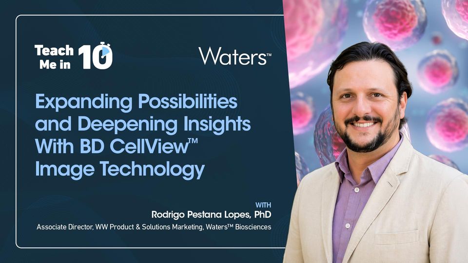
In this Teach Me in 10, join Dr. Rodrigo Pestana Lopes, Associate Director, WW Product & Solutions Marketing at Waters™ Biosciences, to see how imaging-enabled analysis identified 48 functional T-cell subsets compared with 16 using traditional analysis alone.
Watch this episode to discover:
- How spatial information distinguishes cells with similar levels of marker expression but different intracellular localization.
- What 78 imaging-enabled parameters can add to traditional spectral flow cytometry measurements.
- How changes in nuclear morphology and protein localization can reveal additional features of T-cell activation.
Further resources:
- Supercharge Your CAR T Cell Research in Flow Cytometry by Integrating Morphological and Spatial Insights in Real Time
- Remove Blind Spots in Flow Cytometry by Identifying Nuclear Translocation of Transcription Factors With BD CellView™ Image Technology
- Case Study: Revealing Rare and Transient Cell States
- Flyer: Fast and Reliable Real-Time Imaging
Meet the Author

Scientific Content Producer
Interviewing

Associate Director, WW Product & Solutions Marketing