Distinguishing Feature of T Cells in Colorectal Tumors Identified
Complete the form below to unlock access to ALL audio articles.
University of Otago scientists have discovered a way to view the immune cell 'landscape' of bowel cancer tumours, paving the way towards more individualised medicine and treatment for many other diseases in future.
In a paper recently published and featuring on the cover of the Journal of Immunology, the scientists have shown the incredible diversity of immune cells that are inside a colorectal tumour. Immune cells are known to protect against cancer growth and this work provides new information on the types of cells present and how they might be beneficial for the patient.
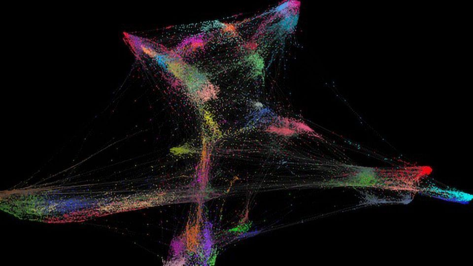
Lead researcher Associate Professor Roslyn Kemp explains they are using a new technology called high-dimensional mass cytometry to identify cells in the tumours of people with bowel cancer.
“It can be thought of as taking a higher resolution ‘photo’ of the inside of the tumour. The photo may reveal new types of cells that may or may not be targetable by drugs, or reveal different composition of immune cell populations in individuals that could be used to predict the course of the disease.”
Associate Professor Kemp says results of their study have shown there is huge diversity in the type of immune cells that infiltrate the tumour, which means that any one, or more likely a combination of many immune cells, could have an effect on patient outcomes.
The technique could be used to study a number of different diseases, she says.
“It demonstrates the use of a new technology to study the immune response in much more detail than other methods currently used, providing new types of information for patients,” Associate Professor Kemp explains.
“It is a step towards personalised medicine, sometimes referred to as precision medicine, since each patient’s tumour could be looked at with this amount of detail.”
The next step is to carry out a similar study with a slightly different technique to further investigate where all the cells are in the tumour and how that might affect cell function and relationships between types of cells and patient data like stage of disease and patient survival.
The research is supported by the Cancer Research Trust, Lotteries Health Research, the University’s School of Biomedical Sciences with Associate Professor Kemp supported by the NZSO Roche Translational Cancer Research Fellowship.
This article has been republished from materials provided by the University of Otago. Note: material may have been edited for length and content. For further information, please contact the cited source.
Reference:
Norton et al. (2019). High-Dimensional Mass Cytometric Analysis Reveals an Increase in Effector Regulatory T Cells as a Distinguishing Feature of Colorectal Tumors. The Journal of Immunology. 202: 1871-1884

