High-Resolution Experiments Reveal How Cells "Eat"
Complete the form below to unlock access to ALL audio articles.
A new study shows how cell membranes curve to create the “mouths” that allow the cells to consume things that surround them.
“Just like our eating habits basically shape anything in our body, the way cells ‘eat’ matters for the health of the cells,” said Comert Kural, associate professor of physics at The Ohio State University and lead author of the study. “And scientists did not, until now, understand the mechanics of how that happened.”
The study, published recently in the journal Developmental Cell, found that the intercellular machinery of a cell assembles into a highly curved basket-like structure that eventually grows into a closed cage. Scientists had previously believed that structure began as a flat lattice.
Membrane curvature is important, Kural said: It controls the formation of the pockets that carry substances into and out of a cell.
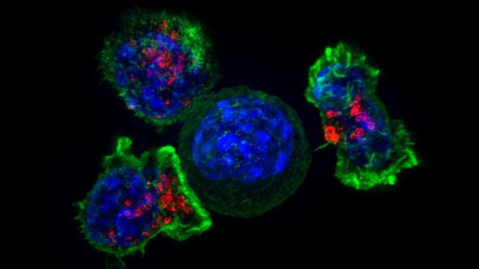
The pockets capture substances around the cell, forming around the extracellular substances, before turning into vesicles – small sacs one-one millionth the size of a red blood cell. Vesicles carry important things for a cell’s health – proteins, for example – into the cell. But they can also be hijacked by pathogens that can infect cells.
But the question of how those pockets formed from membranes that were previously believed to be flat had stymied researchers for nearly 40 years.
“It was a controversy in cellular studies,” Kural said. “And we were able to use super-resolution fluorescence imaging to actually watch these pockets form within live cells, and so we could answer that question of how they are created.
“Simply put, in contrast to the previous studies, we made high-resolution movies of cells instead of taking snapshots,” Kural said. “Our experiments revealed that protein scaffolds start deforming the underlying membrane as soon as they are recruited to the sites of vesicle formation.”
That contrasts with previous hypotheses that the protein scaffolds of a cell had to go through an energy-intensive reorganization in order for the membrane to curve, Kural said.
The way cells consume and expel vesicles plays a key role for living organisms. The process helps clear bad cholesterol from blood; it also transmits neural signals. The process is known to break down in several diseases, including cancer and Alzheimer’s disease.
“Understanding the origin and dynamics of membrane-bound vesicles is important – they can be utilized for delivering drugs for medicinal purposes but, at the same time, hijacked by pathogens such as viruses to enter and infect cells,” Kural said. “Our results matter, not only for our understanding of the fundamentals of life, but also for developing better therapeutic strategies.”
Reference: Willy NM, Ferguson JP, Akatay A, et al. De novo endocytic clathrin coats develop curvature at early stages of their formation. Dev Cell. 2021;56(22):3146-3159.e5. doi: 10.1016/j.devcel.2021.10.019
This article has been republished from the following materials. Note: material may have been edited for length and content. For further information, please contact the cited source.

