New Insights Into the Cells That Host HIV
Complete the form below to unlock access to ALL audio articles.
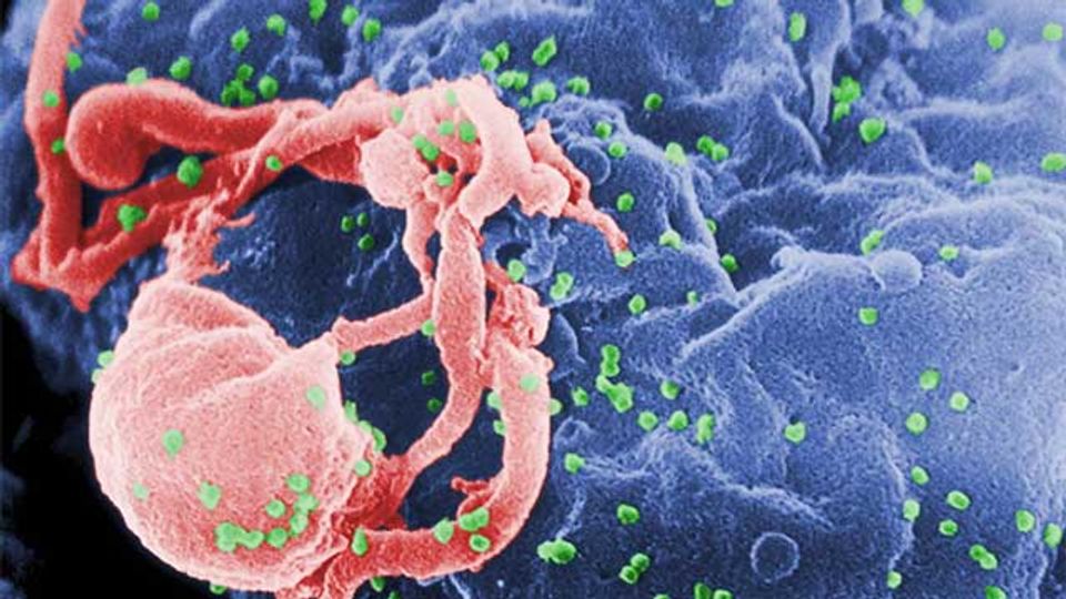
New research sheds light on the lifespans and location of the cells that are responsible for producing HIV, preventing its eradication. Understanding the cells’ dynamics may help scientists develop new ways to reduce their number with the ultimate goal of curing HIV infection.
“When chronically HIV-1 infected individuals are put on potent antiretroviral drug therapy, the amount of virus in their blood decreases,” said Los Alamos National Laboratory Senior Fellow Alan Perelson. He and collaborator David D. Ho, MD, now at Columbia University School of Medicine, found that “the viral decline occurred in two distinct phases, a fast first phase followed by a slow second phase.”
Using a mathematical model of viral infection and treatment developed by Perelson, the team concluded that the two-phase decline reflected the fact that HIV infected two distinct cell populations that produced HIV. One population produced the majority of the virus, but lived only a day or so. The decay of this population according to the model was responsible for the first phase decline of the virus in the blood.
The second cell population, which released virus at a slower rate, lived a matter of weeks while producing virus, and their loss according to the model was responsible for the second phase of viral decay seen in the blood.
Now, in a new paper published in the journal PNAS this week, Robert Siliciano, MD, from Johns Hopkins University School of Medicine and his team working with Perelson and Ruy Ribeiro from the Theoretical Biology and Biophysics Group at Los Alamos National Laboratory searched for these hypothesized cells with different decay rates. Siliciano’s group isolated HIV-infected cells in the blood of 17 people living with HIV on antiretroviral therapy twice a month for the first three months after the initiation of the therapy, and then every month for a year. They found that very few of the short-lived infected cells responsible for the first phase of viral decay were circulating in the blood, suggesting that these cells most likely reside in tissues, such as lymph nodes and the spleen.
Instead, they found cells in the blood that carried an intact HIV genome and which decayed with a half-life of about two weeks. These are presumably the cells responsible for the second phase of viral decay predicted by Perelson and Ho. After about three months on treatment, the remaining infected cells with intact HIV genomes decayed even more slowly, now with a half-life of about 19 months. These cells may become part of the latent-infected cell reservoir, which if therapy is stopped re-seeds the infection and virus then becomes detectable usually in a matter of weeks.
Reference: White JA, Simonetti FR, Beg S, et al. Complex decay dynamics of HIV virions, intact and defective proviruses, and 2LTR circles following initiation of antiretroviral therapy. PNAS. 2022;119(6). doi: 10.1073/pnas.2120326119
This article has been republished from the following materials. Note: material may have been edited for length and content. For further information, please contact the cited source.

