Digital Pathology: The Hidden Infrastructure Behind the Shift to Alternative and Innovative Methods
New approach methodologies are often defined by the models and algorithms at the front end. Their success, however, increasingly depends on something less visible: a validated digital pathology foundation.
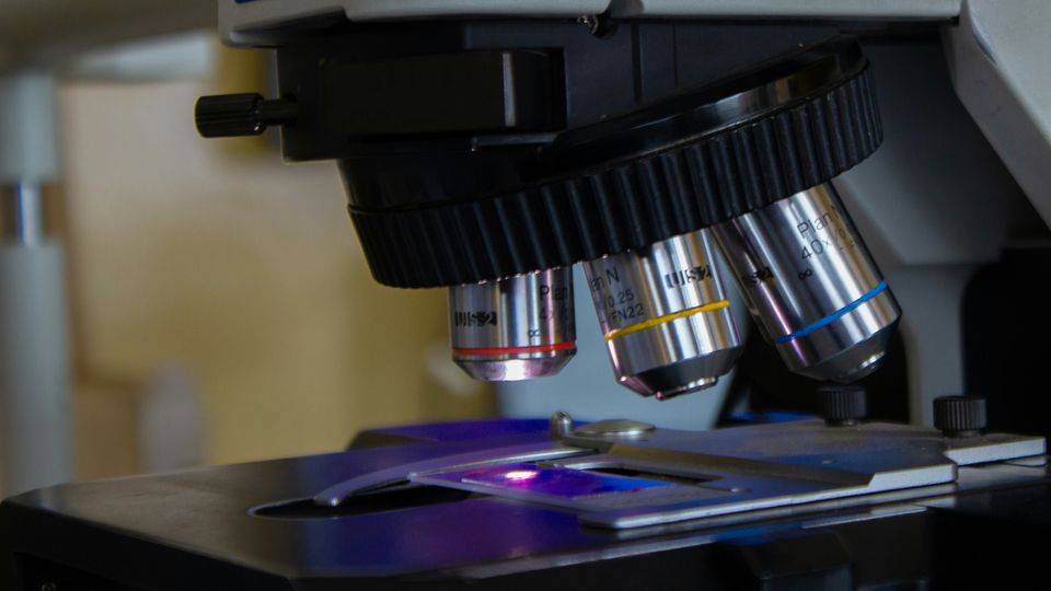
Digital pathology is often presented as an efficiency tool: a way to review slides remotely, share images faster, or reduce the movement of glass slides between laboratories. All of those matter, but this framing also understates its strategic value. Both in clinical and nonclinical research, digital pathology is a core scientific infrastructure that allows increasingly automated image analysis, especially when combined with molecular technologies. Thus, it plays a key role in the transition towards new scientific methodologies.
New approach methodologies (NAMs) span a broad and evolving toolkit, including complex in vitro models, organoids, microphysiological systems, and computational approaches. These technologies are intended to generate more predictive evidence while reducing or refining animal use. Nonetheless, in isolation, new models do not aggregate confidence. Subject-matter experts must be able to connect their outputs with established biological evidence, interpret tissue-level changes, and determine whether an observed signal is biologically meaningful. That is where digital pathology is a game changer.
A tissue slide is more than an image
Histopathology remains one of the most information-rich endpoints in nonclinical safety assessment. A slide captures not only whether a lesion is present, but also its location, distribution, severity, and relationship to surrounding tissue. When that evidence exists only on glass, multidisciplinary exchange at scale is limited.
Whole-slide imaging changes that. A high-quality digital slide can be reviewed alongside study metadata, clinical pathology results, exposure data, and prior findings. It can also become part of a curated reference set that supports cross-study comparison, quantitative image analysis, and the development of computational models.
The value, however, does not come from scanning alone. The image must remain linked to its biological context and its method of study. Tissue identity, staining method, scanner settings, image quality, study design, and pathology terminology all influence interpretation. A useful digital pathology ecosystem therefore requires governance, traceability, standardized metadata, and fit-for-purpose validation—not simply a large image archive.
Virtual control groups need reviewable tissue evidence
Virtual control groups (VCGs) illustrate why this foundation matters as they use carefully selected historical control animals that are matched to a new study and may support a reduction in the number of concurrent control animals. Their purpose is not to replace scientific judgment with a historical average. They are intended to bring relevant prior evidence into the context of a current study and scientific reviews have emphasized their animal-reduction potential given the breadth and consistency of data/tissue collection.
To deliver that value rigorously, pathologists need access to digitized and quality-controlled tissue slides from the selected control animals. This preserves the ability to review the underlying evidence directly, compare morphology across groups, and investigate any unexpected findings.
This is especially important because pathology interpretation is contextual. A rare background lesion, a subtle shift in severity, or a difference in tissue distribution may matter even when summary incidence tables look similar. Digital slides in VCGs keep essential context visible and readily accessible worldwide.
Connecting established pathology with emerging models
Consistent data and tissue collection tied with digital pathology is the infrastructure that helps evaluate and scale up tissue-based alternatives. Organoids, three-dimensional cell cultures, and other complex in vitro models increasingly reproduce elements of tissue architecture. But their added value depends on more than a viability assay or a single molecular marker. Subject-matter experts still need to judge whether the model develops the expected morphology, how it responds to injury, and whether its signals are biologically relevant.
Pathologists are well placed to make those connections. By applying consistent imaging and assessment approaches across conventional histopathology and complex in vitro systems, digital pathology creates a common visual language. When supported by AI tools, image analyses can become faster and/or more quantitative. Further, digital image analysis helps define where and when a model is representative, and which scientific questions it can answer reliably.
That bridge is essential for qualification, as confidence in new methods will not come from demonstrating novelty; it will come from showing reproducibility, biological relevance, and a clearly defined context of use.
From morphology to mechanisms and greater translatability
Digital pathology also provides the spatial anchor needed to integrate morphology with multiomics. Transcriptomics, proteomics, and other molecular methods can reveal treatment-associated pathways, but it is spatial approaches that map those signals within a tissue and to the cell populations involved.
This is particularly valuable when standard histopathology identifies a finding but cannot fully explain its mechanism or relevance to humans. Linking a molecular signature to a specific microscopic change can strengthen the weight of evidence and guide decisions about dose response, reversibility, or follow-up investigation. In some cases (e.g., gene therapies and oligonucleotide-based or antibody-drug conjugate treatments), molecular safety biomarkers and mechanistic pharmacology signals may require multiomic readouts in order to achieve appropriate resolution.
Advances in methods for formalin-fixed, paraffin-embedded tissue are expanding the potential to generate molecular insight from archived and routine specimens. This creates an opportunity to obtain more value from tissue that has already been collected, while targeted prospective fresh or frozen material sampling can be used in parallel when still scientifically necessary. The goal is not to apply multiomics to every study. It is to select the right method when the biological question warrants greater resolution, making digital pathology indispensable.
Artificial intelligence depends on human expertise
Artificial intelligence (AI) can extend these capabilities by detecting patterns, quantifying features, and prioritizing regions for review across large image collections. But the pathologist's role remains crucial in defining the biological question, curating representative data, reviewing morphology, and determining the scientific meaning of a model’s output.
The quality of an algorithm is constrained by the quality and representativeness of the images and annotations used in its development. Differences in tissue processing, staining, scanners, or study populations can introduce unintended variation. Validated workflows, ongoing performance monitoring, and human oversight are therefore central to responsible adoption.
Regulatory expectations are reinforcing this point. The US Food and Drug Administration’s guidance on whole-slide imaging in GLP-compliant nonclinical toxicology studies addresses written procedures, system validation, documentation, image management, and retention. These controls are not barriers to toxicology research—they are the very mechanisms that make digital evidence dependable enough to support innovation.
For NAMs, infrastructure comes first
The shift toward NAMs is sometimes described as a move away from established nonclinical science. In practice, progress will depend on connecting new tools with the strongest evidence and expertise already available.
Digital pathology makes those connections more efficient. It enables historical tissue to be reused responsibly, gives VCGs a reviewable morphological foundation, supports the qualification of complex in vitro models, and links molecular signals with tissue context. It also creates the controlled data environment required for AI-enabled analysis.
The organizations best positioned for the next phase of nonclinical innovation will not be those that simply accumulate the most images or deploy the most algorithms. They will be those that build trusted, interoperable pathology ecosystems in which data, tissue evidence, and expert judgment reinforce one another. Digital pathology may be the less visible part of the NAM transition, but it is one of the foundations on which that transition will stand.
 Add Technology Networks as a preferred Google source to see more of our trusted coverage.