Phagocytosis vs Pinocytosis: Definition and Function

Complete the form below to unlock access to ALL audio articles.
Phagocytosis and pinocytosis are biological processes wherein a cell senses and engulfs nearby material. These processes enable cells to intake substances that can’t easily pass through the cell membrane. However, phagocytosis and pinocytosis are forms of endocytosis that differ in several ways including how they intake material and the type of material they engulf. In this guide, we explore the basics and key differences between phagocytosis vs pinocytosis in more detail.
What is phagocytosis?
Phagocytosis is a specialized process by which cells engulf relatively large, solid material. These particles, generally larger than 0.5 μm in diameter, may include apoptotic cells or foreign substances. Unicellular organisms such as amoebas use phagocytosis to acquire nutrition while cell types of multicellular organisms use this universal process for preventative functions such as tissue homeostasis. Among these cells, certain types are capable of greater efficiency in the phagocytic process, such as monocytes or macriophages. Other, like epithelial cells, are capable of phagocytosis, but at a less efficient rate.Phagocytosis function
The cellular processes of phagocytosis consist of four distinct phases that include 1) the detection of target material, 2) activation of phagocytosis, 3) the formation and 4) maturation of the phagosome.

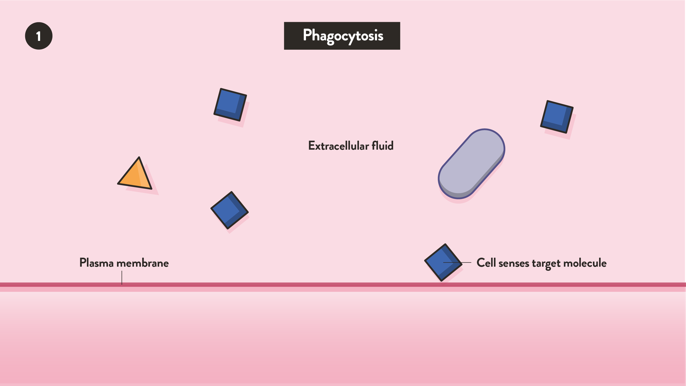
Image 1: Phagocytosis: The detection of target material
The detection of target material begins when a cell senses target material on its surface (Image 1). Most cell types have a dedicated set of cell surface receptors that recognize various substances. For example, immune cells can sense invasive microbes with receptors that recognize pathogen-associated markers. After the target material has been detected, the cell surface receptor initiates a series of signals that prepare the cell for phagocytosis.

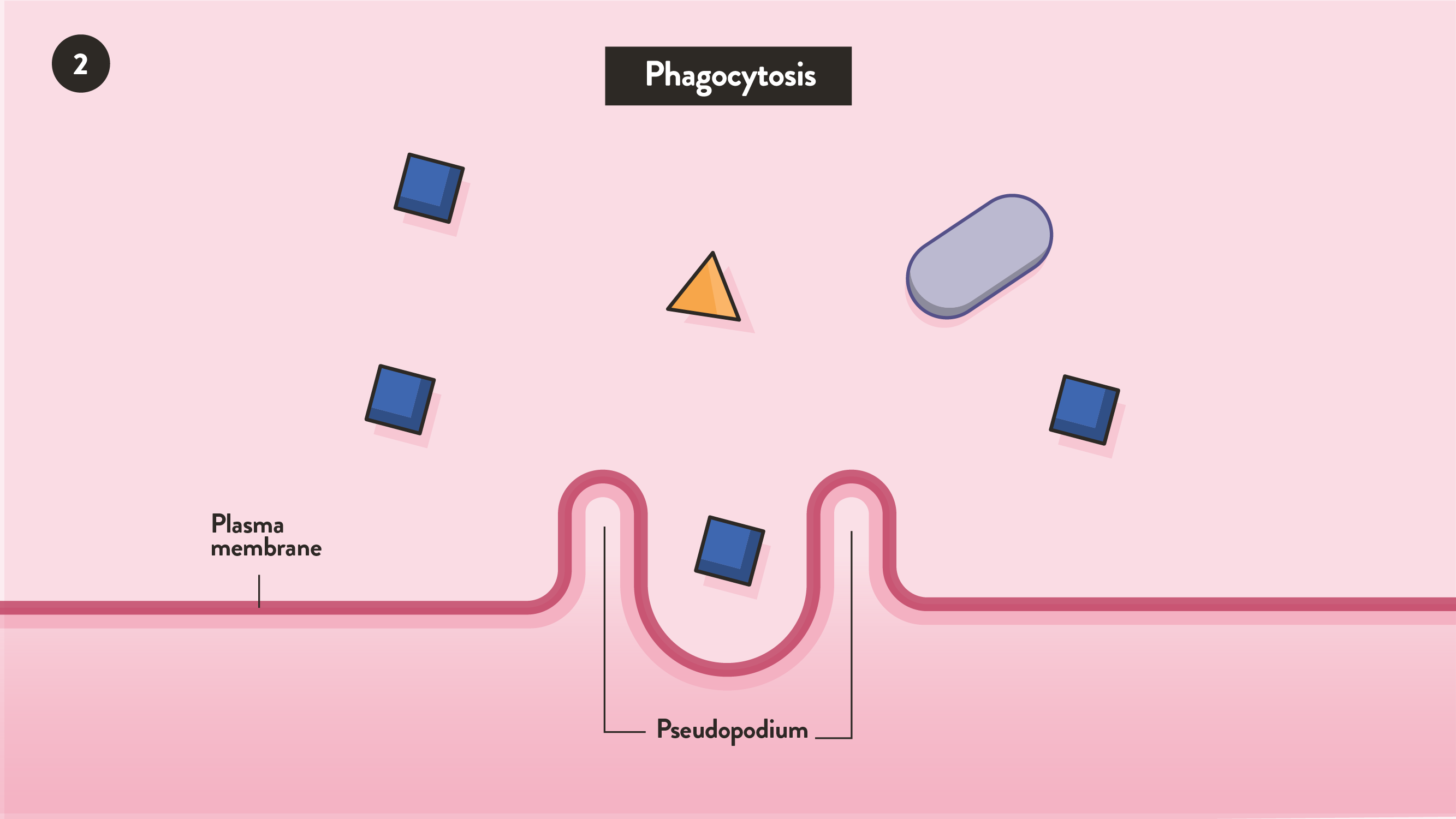
Activation of phagocytosis causes the cell membrane area in contact with the target material to rearrange into a cavity called the phagocytic cup (Image 2). This process begins when the material-stimulated receptors send molecular signals that prepare the cell for target material intake.

The formation of the phagosome is the third phase of phagocytosis that involves the complete internalization and closure of the phagocytic cup (Image 3). Once the phagocytic cup fully covers and internalizes into a cellular compartment, the newly formed phagosome can begin its maturation process.

Image 4: Phagosome maturation. Here, the phagosome has merged with a lysosome, forming a phagolysosome.
Maturation of the phagosome is the final phase of phagocytosis (Image 4). Phagosome maturation consists of a series of material handoffs between the original phagosome and cellular compartments specialized in digestion. The early stages of phagosome maturation include a combination of fusion and fission events that prepare the cellular membrane of the maturing phagosome for material digestion. During the final stages of phagocytosis, the internal environment of the mature phagosome becomes more acidic and reactive, which aids the digestion of target material such as microorganisms.
What is pinocytosis?
While phagocytosis involves the ingestion of solid material, pinocytosis is the ingestion of surrounding fluid(s). This type of endocytosis allows a cell to engulf dissolved substances that bind to the cell membrane prior to internalization. Unlike phagocytosis, pinocytosis is a “drinking” mechanism wherein a cell actively engulfs external fluids over time. Even though pinocytosis differs from other forms of receptor-mediated endocytosis, these terms overlap with one another due to their similarities.
Pinocytosis function

In humans, pinocytosis primarily occurs when cells absorb nutritional or waste droplets suspended in external fluid. The nutritional molecules that can activate pinocytosis include fats, sugars, proteins, ions or other small molecules. This process begins when a soluble substrate binds to the surface of a cell (image B1)


This pinosome continues to pinch off from the cell membrane (Image 6B) within the cell until it is fully internalized (Image 7).

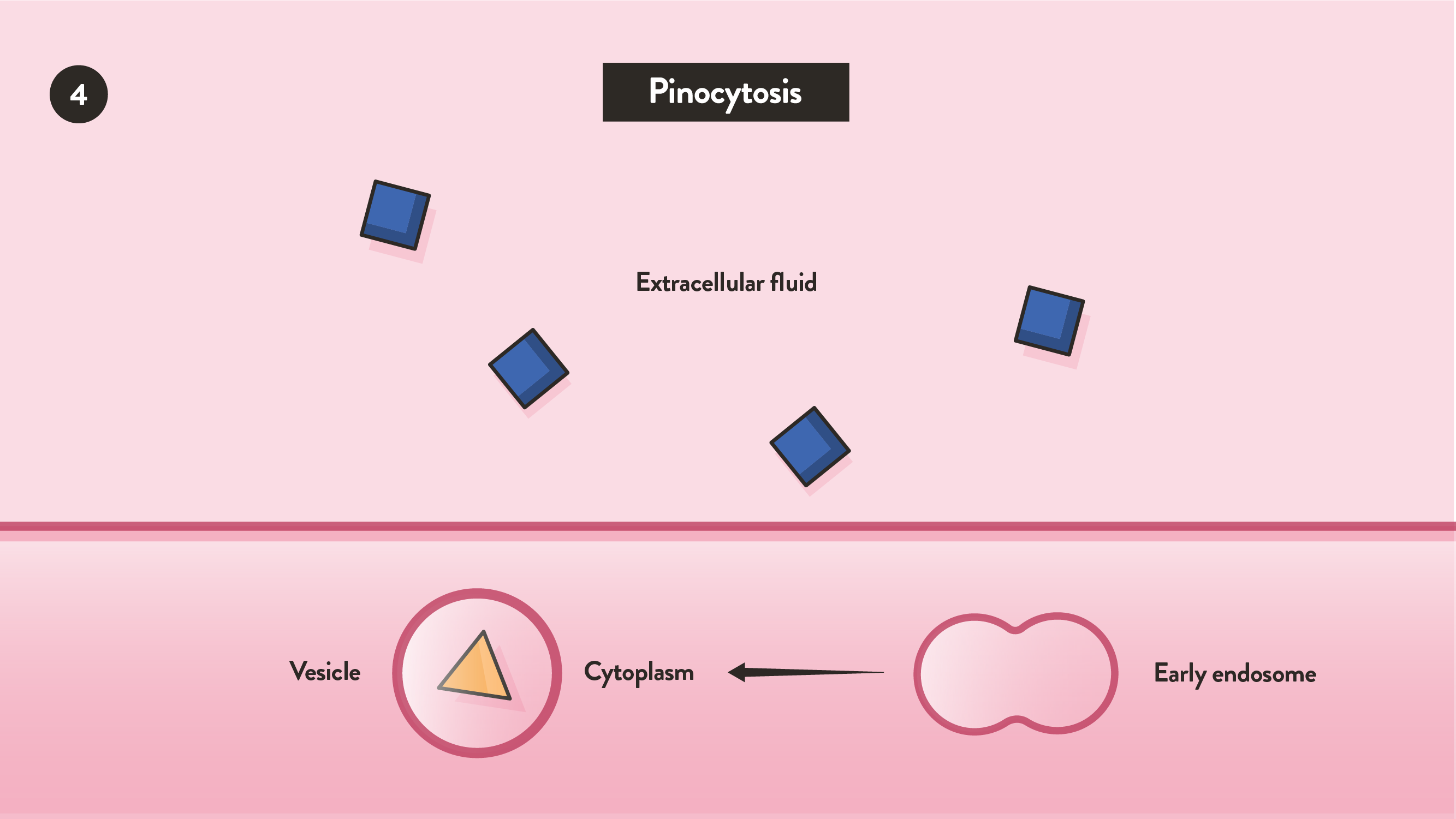
Once the cell fully internalizes this cellular pocket, the newly formed compartment can swap its material with specialized vesicles that can break down substrate into smaller molecules (Image 8).
The size of these vesicles during the intake of fluid material can establish whether a cell performs macropinocytosis vs micropinocytosis. Macropinocytosis is a form of non-specific endocytosis that is common for all cell types. This process includes the intake of fundamental nutrients such as amino acids, carbohydrates and fats.
Micropinocytosis involves the ingestion of fluid material by small vesicles that are typically no larger than 0.1 micron. This process is typically initiated by cell surface lipid domain once they bind to their specific target molecules.1
Phagocytosis vs pinocytosis: chart
|
| Phagocytosis | Pinocytosis |
| Definition | Cellular intake of solid material | Cellular intake of fluid |
| Role | Immune response, nutrient uptake | Immune surveillance, nutrient uptake |
| Cell Types | Most commonly immune cells | Almost all cell types |
| Example | Macrophages | Skin cells |
Examples of phagocytes and phagocytic cells
Phagocytes are a specialized group of cells that perform phagocytosis efficiently. This group includes immune cells such as macrophages, neutrophils and monocytes. Each type of phagocyte has a set of cell surface receptors that can initiate phagocytosis whenever they sense a target particle or pathogen.
Opsonization in phagocytosis
Opsonization is an immune process where phagocytes bind to foreign molecules that are linked to host-derived proteins such as antibodies. This process involves the binding of free-floating antibodies to foreign materials so that these potential threats can be identified by the immune system. The tail of each antibody has a segment called the Fc region. This region specifically binds to Fc cell surface proteins found on the surface of phagocytic cells. Opsonization can also occur via the complement system, where a fragment of the system, C3b, acts as a binding site for phagocytes.
Opsonization is completed when a phagocyte identifies the foreign molecules of interest and initiates phagocytosis.


