ASCB: A CELLebration of Cell Biology

Complete the form below to unlock access to ALL audio articles.
Early December saw an unseasonably cold San Diego hosting the American Society of Cell Biology’s annual conference. Most instrument vendors focused on visualization as a major theme, especially using variants of super-resolution microscopy, while antibody companies were all keen to address the negative stories that have emerged in 2015 around the lack of consistency and unreliability of antibodies in research making studies unreplicable
Technology Networks met with a range of companies from big name instrument vendors to small start-ups, but for three notable exceptions there was not much innovation on display, instead new applications of existing technologies, and developments of existing products.
It was Carl Zeiss and its partner arivis who created the biggest stir with their demonstration of virtual reality (using the famed Oculus Rift headset) to bring to life 3D cell data, although a prototype at this stage, a new product is being considered for release in 2016. Bruker unveiled what it calls the world’s only quantitative super-resolution microscope, start-up AIM Biotech (spun out of MIT) with its 3D cell culture chip for modelling complex biological systems. The all-plastic chip contains interlinked channels filled with gel in which cells can grow. AIM provided a nice application of the system for modelling the metastasis of breast cancer into bone marrow. Here users can create 3D blood vessels, bone cells and stem cells in the gel to mimic the living marrow of a bone, and were able to model the spread of cancer cells into the marrow microenvironment. The various cell types behaved as they normally would in a clinical setting.
Super-Resolution Microscopy:
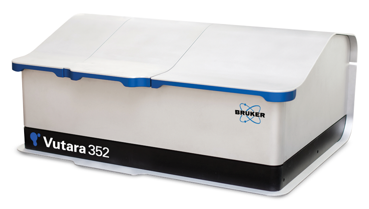
Bruker launched the Vutara 352, the fastest super-resolution, single-molecule localization (SML) microscope commercially available. The Vutara 352 offers speed, imaging depth and resolution to deliver significant advantages over competing technologies, in addition to incorporation of quantitative analysis modules into the workflow based instrument software. For the first time, this represents an entirely new dimension of functionality in super-resolution microscopy, including the ability to perform pair-correlation, co-location, cluster, and live-cell analysis along with super resolution microscopy.
![]()
The Vutara 352 is now also compatible with Bruker’s Opterra multi-point confocal scanner, creating a unique combination of super-resolution and confocal capabilities, where both instruments are designed for high-speed imaging. This enables the visualization of large-scale, high-resolution structural context in correlation with super-resolution imaging in even the most challenging biological preparations.
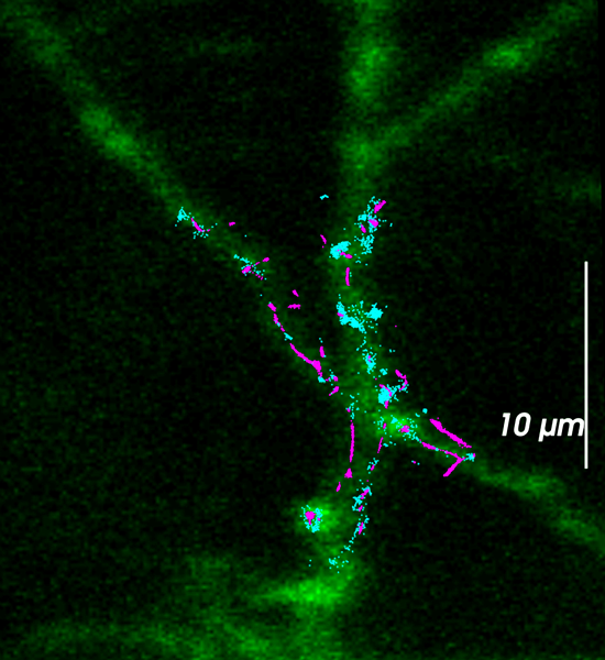
“The addition of the Opterra in the next generation Vutara provides a high resolution optical sectioning mode of operation for sample navigation that provides a clearer picture of areas of interest for super resolution acquisition, Jeff Stuckey explained. “The post-acquisition correlative overlay provides contextual information for visualization and analysis of super resolution data”.
Confocal Microscopy:
ZEISS showcased its LSM 880 and LSM 800 confocal microscopes with Airyscan technology, providing 1.7 fold increase in resolution over conventional confocal instruments to provide high signal-to-noise data from live cells or other weakly labelled samples. With Airyscan, instead of throwing light away at the pinhole, a 32 channel area detector collects all light of an Airy pattern simultaneously. Each detector element functions as a single, very small pinhole. Knowing the beampath and the spatial distribution of each Airy pattern enables a very light-efficient imaging: enabling the use of all the photons collected. Duncan McMillan (Director of Product Marketing) explained that confocal is a long established technique that is preferred over some of the new superresolution techniques, ‘by adding Airyscan technology, it provides users with the power of superresolution microscopy in an easy to use format, with little barrier to entry.’
Olympus highlighted its compact, easy to use, spinning-disk laser confocal microscope (CV1000), which is designed for long-term time-lapse imaging. The compact solution provides the same high-end image quality of a traditional confocal system, but without the need for a highly trained imaging expert or a darkroom. Olympus’ SD-OSR super resolution microscope is a live cell imaging that produces 120 nm, super resolved, 4-megapixel images at 3 frames / second. The SD-OSR requires no special sample preparation and achieves dynamic super resolution from resolution limited Nipkow spinning disk illumination of the sample
Microscope Cameras:
ZEISS introduced two new digital microscope cameras: ZEISS Axiocam 702 mono and ZEISS Axiocam 512 color. ZEISS Axiocam 702 mono is a microscope camera with a scientific CMOS sensor. Users benefit from low read noise, excellent low light sensitivity and high speed for live cell imaging and acquisition of fast processes. ZEISS Axiocam 512 color allows acquisition of large sample areas in one high resolution, true color image.
Hamamatsu updated its ORCA-Flash4.0 with V2 to provide an increase in the ability to detect photons. The enhanced QE (Quantum Efficiency) provides the possibility of detecting faint signals, or, for moderately dim samples, that the ideal image is achievable with shorter exposure times.
Photometrics introduced its new PRIME sCMOS camera. Photometrics Prime is the first intelligent scientific CMOS camera to incorporate FPGA-based Embedded Signal Processing™ engine (ESP). ESP enables advanced real-time processing features: PrimeEnhance quantitatively increases the Signal to Noise Ratio by 3X-5X, increasing the clarity and quality of images. PrimeLocate dynamically evaluates acquired images and reduces the surplus of data generated during high speed super resolution imaging.
QImaging unveiled a new range of CCD cameras: Retiga R Series, which provide solutions from routine imaging and documentation to advanced live cell imaging.
Cell Culture Technology:
Olympus’ CKX53 inverted microscope provides improved image capabilities and ergonomics, for enhanced performance and a comfortable workflow for a variety of cell culture needs such as live cell observation, cell sampling and handling, image capture, and fluorescence observation. A second product highlighted is the VS120. VS120-S6-W slide loader system allows manual loading of one and six standard slides, respectively, along with any associated meta-data. Designed for high throughput research and pathology, the VS120-L100-W system features a highly dependable, robustly designed slide loader for up to 100 slides.
Bio-Rad hailed its S3E Cell Sorter to be the first truly walk-away automated cell sorter. Real-time monitoring and smart features make cell sorting easier and accessible to both novices and experts alike. This compact system is equipped with either one or two lasers and up to four fluorescence detectors plus forward- and side-scatter detectors.
MilliporeSigma highlighted the Muse Cell Analyzer a robust, compact instrument for automated cell analysis and its update to the Amicon Stirred Cells. Amicon stirred cells are ideal for finely separating macrosolutes (DNA, RNA, protein, etc.) in large-volume samples. They are also reliable tools for membrane analysis, because they allow users to insert membranes of choice. The upgrade brings improved performance and enhanced design and ergonomics.
Ibidi showcased a new slide for imaging featuring a unique yet proven polymer base. The µ-Slide for correlative light and electron microscopy (CLEM), and perfusion-based assays, enables cell growth with better performance than traditional glass.
Gene Editing:
Edit-R CRIPSR-Cas9 Genome Engineering tools provide high-confidence gene editing. The Dharmacon Edit-R CRISPR-Cas9 platform from GE Healthcare provides pre-designed, ready-to-use DNA and RNA components to enable rapid and highly functional gene editing experiments.
Horizon highlighted how using CRISP-R Cas9 produces robust pipelines for cell-line models. This promotes efficient and robust pipelines to produce knock-out cell lines, within a week at a good price.
Antibodies and antibody technology:
Antibodies have been found to be unreliable reagents resulting in poor specificity, sensitivity and consistency. This has resulted in false findings and wasted efforts. Frustration with research antibodies has been developing for years, but now there is a big interest to improve current issues.
One World Lab has developed an online platform to help researchers decide which antibodies to choose, with the intention of acting as a bridge for the flow of information between manufacturers and researchers. The company also provides antibodies that are traceable to the original manufacturer and low-cost ‘trial size’ antibody batches. This enables researchers to compare antibodies side by side in their labs, allowing them to select the best antibody for the application.
Miltenyi Biotec highlighted the significance of recombinant antibody production for enhanced reproducibility. Recombinant antibodies are different to traditional mono- and polyclonals because they are defined by a genetic sequence on a recombinant expression vector. Commercial production of recombinant antibodies, however, requires a significant change in production technology, resulting in a lack of product breadth on the market. Miltenyi’s response to this market gap is their portfolio of REAfinity™ Antibodies which currently includes over 4,000 antibodies and is rapidly expanding in number, thereby providing researchers with consistent antibodies for enhanced reproducibility of scientific data.
Thermo Fisher Scientific was keen to highlight its antibody selection tool which showcases more than 40,000 of its Invitrogen™ antibodies. Antibody assay results are validated internally by a team of more than 100 scientists and links are provided to thousands of citations worldwide, to provide confidence in antibody choice."
Software:
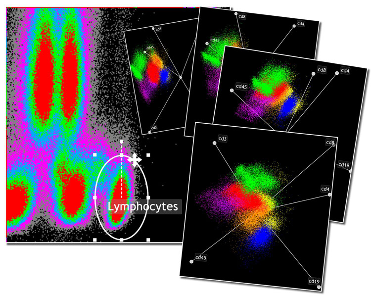
Beckman Coulter launched Kaluza 1.5 software for flow cytometry. Kaluza aims to provide a greater understanding of the data generated by modern cytometers. Kaluza employs technology that allows for real time analysis of high content files allowing you to spend your valuable time on discovery. V1.5 includes added visualization enhancements such as improved overlay plots (histogram, density and dot) and better smoothing for histograms. Panning and zoom allow you to manipulate your data with ease.