Top 10 Cell Science News Stories of 2018


Featuring a solid mix of imaging, cell and animal models, 3D printing and CRISPR, here is a wrap-up of the most-read cell science news stories we covered in 2018.
While it's a bit late to put your orders in this year, now is your chance to have your say for 2019. What cell biology topics would you like to hear about next year? Drop us a line!
Merry Christmas and Happy New Year all!
1. Possible Culprit of Fibromyalgia Found: Microglial Activation
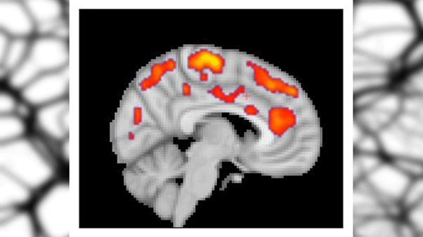 This combined MR/PET image highlights areas of the brain in which patients with fibromyalgia were found to have increased glial activation, compared with unaffected control volunteers. Image credit: Marco Loggia, PhD, Martinos Center for Biomedical Imaging, Massachusetts General Hospital)
University/Institute: Massachusetts General Hospital, Karolinska Institutet
Link to paper, published in: Brain, Behavior and Immunity
In a nutshell: 31 fibromyalgia patients and 27 healthy controls were scanned using Position Emission Tomography, to assess brain glial activation. Fibromyalgia patients exhibited elevated cortical levels of [11C]PBR28 signal, which binds to a protein upregulated in activated microglia and astrocytes.
Author insights: “The activation of glial cells we observed in our studies releases inflammatory mediators that are thought to sensitize pain pathways and contribute to symptoms such as fatigue.” -Marco Loggia, assistant professor of Radiology at Harvard Medical School.
2) Fasting Boosts Stem Cells’ Regenerative Capacity in Mice

Image credit: Pixabay
University/institute: Massachusetts Institute of Technology
Link to paper, published in: Cell Stem Cell
In a nutshell: A 24 hour fast was shown to improve intestinal stem cell function in mice by inducing a fatty acid oxidation program. Pharmacological activation of this program mimics many effects of fasting. This study represents a potential strategy for enhancing intestinal regeneration.
Author insights: “Interestingly, switching these cells to fatty acid oxidation enhanced their function significantly. Pharmacological targeting of this pathway may provide a therapeutic opportunity to improve tissue homeostasis in age-associated pathologies.” -David Sabatini, MIT professor and senior author.
3) 2 Clinically-approved Antibiotics Destroy Senescent Cells
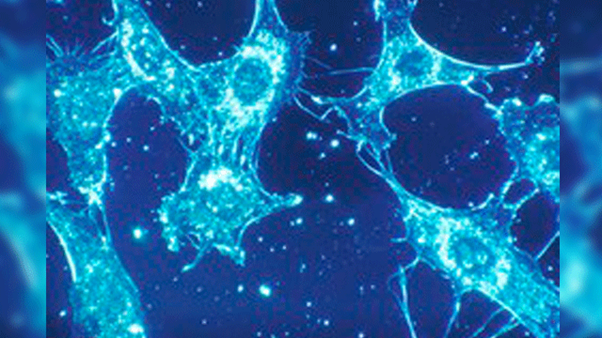 Normal cells of human connective tissue in culture. At a magnification of 500x, the cells were illuminated by darkfield amplified contrast technique. Image credit: National Cancer Institute
University/institute: University of Salford
Link to paper, published in: Aging
In a nutshell: With the goal of repurposing FDA-approved antibiotics, antibiotics were screened for their ability to selectively induce death of senescent cells. Two candidates were identified, one of which is commonly prescribed to cystic fibrosis patients — raising questions regarding the mode of action of this mild antibiotic.
Author insights: “Originally, the thinking was that Azithromycin is killing harmful bacteria in cystic fibrosis patients – but our tests now shed a new light on what might be actually going on. Our new interpretation is that the antibiotic is probably eliminating the 'inflammatory' fibroblasts, in other words, the senescent cells that are normally associated with agiing. If that is the case, then we may have unearthed a very inexpensive and readily available method of eliminating aging cells that are toxic to the body.” -Professor Michael P. Lisanti.
4) Neuronal Stem Cells 3D-printed onto Potential Implant
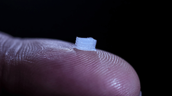
University of Minnesota researchers developed a prototype of a 3D-printed device with living cells that could help spinal cord patients restore some function. The size of the device could be custom-printed to fit each patient’s spinal cord. The patient’s own cells would be printed on the guide to avoid rejection in the body. Image credit: University of Minnesota
University/institute: University of Minnesota
Link to paper, published in: Advanced Functional Materials
In a nutshell: Induced pluripotent stem cell-derived spinal neuronal progenitor cells and oligodendrocyte progenitor cells were placed within 3D-printed biocompatible scaffolds made of silicone. The cells then differentiated and extended axons throughout microscale scaffold channels — an exciting observation for this team who is working towards developing a treatment to help people with spinal cord injuries.
Author insights: “3D printing such delicate cells was very difficult,” McAlpine said. “The hard part is keeping the cells happy and alive. We tested several different recipes in the printing process. The fact that we were able to keep about 75 percent of the cells alive during the 3D-printing process and then have them turn into healthy neurons is pretty amazing.” -Michael McAlpine, Ph.D.
“We’ve found that relaying any signals across the injury could improve functions for the patients,” Parr said. “There’s a perception that people with spinal cord injuries will only be happy if they can walk again. In reality, most want simple things like bladder control or to be able to stop uncontrollable movements of their legs. These simple improvements in function could greatly improve their lives.” -Ann Parr, M.D., Ph.D.
5) A Surprising New Cell Structure Discovery
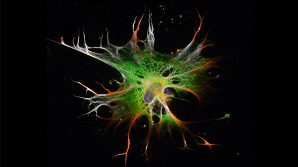
 3D-projection of a cancer cell that is about to undergo cell division and adheres to the substrate with reticular adhesions. Blue: Chromatin/DNA; Red: Cell membrane; Green/Yellow in the bottom of the cell: Reticular adhesions. Confocal microscopy photo by John Lock
University/institute: Karolinska Institutet
Link to paper, published in: Nature Cell Biology
In a nutshell: A distinct class of cell-matrix adhesions have been identified, which are morphologically and dynamically distinct from classical focal adhesions. These ‘reticular adhesions’ maintain cell-extracellular matrix attachment sites during cell division.
Author insights: “It’s incredibly surprising that there’s a new cell structure left to discover in 2018… The existence of this type of adhesion complex has completely passed us by.” -Principal investigator Staffan Strömblad, professor at the Department of Biosciences and Nutrition, Karolinska Institutet.
6) How Your Brain Experiences Time
 A Neural Clock for Time in Experience and Memories. Infographic: Kolbjørn Skarpnes & Rita Elmkvist Nilsen / NTNU & Kavli Institute for Systems Neuroscience
University/institute: Kavli Institute for Systems Neuroscience Centre for Neural Computation
Link to paper, published in: Nature
In a nutshell: Norwegian researchers sought to identify structures responsible for the encoding of temporal information in rats. The conclusion was that populations of lateral entorhinal cortex neurons represent time through the encoding of experience, and that this information may be integrated with spatial inputs from the medial entorhinal cortex in the hippocampus — allowing the hippocampus to store a unified representation of what, where and when.
Author insights: “I believe distributed networks and the combination of structures of activity may deserve more attention in the future. With this work, we have found an area with activity so strongly relating to the time of an event or experience, it may open up a whole new research field.” -Professor Edvard I. Moser.
7) Your Spinal Discs Could Be Replaceable
 Image credit: Pixabay
University/institute: Penn Medicine
Link to paper, published in: Science Translational Medicine
In a nutshell: Penn Medicine researchers sought to develop a treatment for intervertebral disc degeneration, that addresses the underlying cause of degeneration (unlike disc fusion surgery). Tissue-based discs were engineered by sandwiching hydrogel and polymer materials, seeded with cartilage or stem cells between polymer endplates. The engineered discs were implanted into rodents and goats and integrated with native discs in both species. Structure and ‘near-native’ mechanical properties were maintained in both species (rodents: five months after implantation; goats: two months after implantation).
Author insights: “I think it’s really exciting that we have come this far, from the rat tail all the way up to human-sized implants… When you look at the success in the literature from mechanical devices, I think there is a very good reason to be optimistic that we could reach that same success, if not exceed it with the engineered discs.” - Harvey E. Smith, MD, associate professor of Orthopaedic Surgery and Neurosurgery at the Perelman School of Medicine and Staff Surgeon at the CMC VAMC.
8) Tiny Mitochondria Stimulate Brain Cell Connections
 Left: a neuron with all its synapses shown in green. Right: the same neuron with its mitochondria shown in white. Image credit: Tommy Lewis/Polleux lab/Columbia's Zuckerman Institute
University/institute: Columbia University in the City of New York
Link to paper, published in: Nature Communications
In a nutshell: In neurons, mitochondria length varies; cortical, long-range projecting pyramidal neurons have long mitochondria, whereas mitochondria in axons are uniformly short. The functional significance of this ‘shortness’ was explored using in vitro (human embryonic kidney cells) and animal models (mice). A receptor called ‘MFF’ (mitochondrial fission factor) was identified as a key determinant of mitochondria size. By downregulating MFF, it became clear that MFF also affects mitochondria Ca2+ uptake — thereby identifying a novel mechanism controlling neurotransmitter release and axonal branching.
Author insights: “These axonal mitochondria are unlike any mitochondria anywhere else in the body — they are even different than those found in other parts of the neuron. This begged the question: Does this small size serve a function? Not delving into the inner workings of the brain at this level of detail would be akin to trying to understand how a car works just by watching it move along a highway. You have to open up the car’s hood and take a close look at all its parts.” -Dr. Polleux, Professor of neuroscience at Columbia University Irving Medical Center.
9) Astrocytes Derived from Stem Cells in Record Time
 Astrocytes grown from embryonic stem cells. Image credit: Isaac Canals
University/institute: Lunds University
Link to paper, published in: Nature Methods
In a nutshell: Overexpressing the transcription factors SOX9 and NFIB in human pluripotent stem cells was shown to rapidly and efficiently yield homogenous populations of astrocytes. The new method reduces the time it takes to produce the cells from months to two weeks. Obtaining adult human astrocytes is key for advancing research on the role of astrocytes in health and disease.
Author insights: "By combining CRISPR-Cas9 in this way and our method of rapidly cultivating human astrocytes, improved opportunities are available to investigate the role of astrocytes in different neurological diseases," -Henrik Ahlenius.
10) Honeybee Protein Keeps Stem Cells Youthful
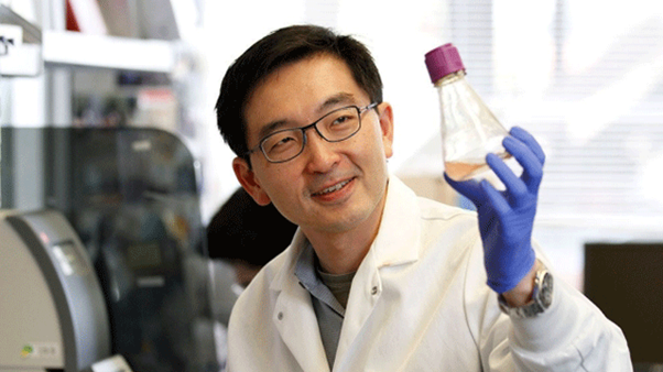 Kevin Wang holds a flask of cells that have been engineered to produce the Regina protein, a mammalian protein similar in structure to the active component of honeybee royal jelly. Photo credit: Paul Sakuma
University/Institute: Stanford University
Link to paper, published in: Nature Communications
In a nutshell: A major protein component of ‘royal jelly,’ i.e. the gelatinous gloop produced by honey bees and fed to larvae/potential queen bees, maintains pluripotency of mouse embryonic stem cells. ‘Royalactin’ activates a ‘ground-state’ pluripotency-like gene network, and induces a naïve-like state in the cells.
Author insights: “In folklore, royal jelly is kind of like a super-medicine, particularly in Asia and Europe… but the DNA sequence of royalactin, the active component in the jelly, is unique to honeybees. Now, we’ve identified a structurally similar mammalian protein that can maintain stem cell pluripotency… It’s fascinating. Our experiments imply Regina is an important molecule governing pluripotency and the production of progenitor cells that give rise to the tissues of the embryo. We’ve connected something mythical to something real.” -assistant professor of dermatology Kevin Wang, MD, PhD, “


