Miltenyi Biotec Launches Fully Automated Light Sheet Microscope
Complete the form below to unlock access to ALL audio articles.
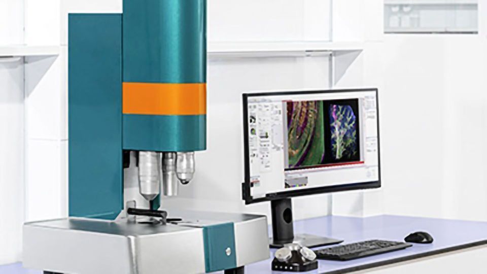
Miltenyi Biotec has announced the UltraMicroscope Blaze, the world’s first fully automated light sheet microscope. The new instrument enables users to image an entire mouse or multiple samples at subcellular resolution in 3D. Automated light sheet imaging of multiple samples together holds the potential to accelerate basic and biomedical research in the field of immuno-oncology and neuroscience.
Offering a new perspective on organisms, on how they are structured, and how they work, the UltraMicroscope Blaze’s technology, together with the specially developed MI Plan objective series, is designed to allow imaging across scales, from whole transparent mice down to subcellular structures.
Benefits of the new UltraMicroscope Blaze include:
- Full automation: The UltraMicroscope Blaze enables automated sample release and smooth switching between different objectives and magnification lenses while keeping images sharp with the autofocus feature.
- Imaging of a large sample or multiple samples: The large sample chamber can either host an entire cleared mouse model or else allows automated imaging of multiple samples. These samples can then be imaged sequentially without the need for manual intervention or monitoring.
- Cutting-edge optics: Optimized illumination guarantees homogeneous fluorescence excitation, and the specially developed MI Plan objective series delivers unprecedented image quality. In combination with the automated magnification changer, these objectives can achieve a total magnification from 0.6× to 30× for subcellular imaging. Additionally, the objectives are compatible with all available clearing solutions, providing full experimental flexibility to researchers.
Applications of the UltraMicroscope Blaze
It is hoped that the field of oncology will benefit from this new technology. Ali Ertürk, Director at the iTERM, Helmholtz Zentrum München, Germany, and his team are among the first users of the instrument. “The UltraMicroscope Blaze allows us to see every single cancer metastasis in whole bodies of transparent mice, and we can also see if drugs are targeting all those tiny micro-metastases. The UltraMicroscope Blaze will be a powerful tool for drug development in oncology,” Ertürk explained. In the field of immuno-oncology, the UltraMicroscope Blaze could open up completely new horizons, for example by allowing 3D imaging of invading tumor-infiltrating lymphocytes, localizing CAR T cells in entire organisms, or analyzing the tumor microenvironment at high resolution.
For neuroscientists, the UltraMicroscope Blaze offers the option to generate 3D images of whole brains in unprecedented detail, facilitating studies on the pathology of Alzheimer’s and Parkinson’s disease in mouse models, for example.
Clearing of samples – the key to high-resolution 3D imaging
To get the best imaging results, appropriate sample preparation is essential when performing light sheet microscopy. Miltenyi Biotec offers a complete portfolio of validated antibodies and antibody-fluorochrome conjugates, and the easy-to-use new MACS® Clearing Kit, fully supporting the light sheet imaging workflow.
The light sheet microscopy technology
Available volumetric imaging techniques, such as magnetic resonance imaging (MRI), can image entire organs and animals but lack the ability to detect small metastases. The UltraMicroscope Blaze overcomes this limitation and bridges the gap between high-resolution imaging of tissue sections and low-resolution imaging of whole animals. The system uses light sheet illumination that is perpendicular to the axis of observation. This decoupling of illumination and detection allows the microscope to operate as a virtual microtome that is able to visualize large transparent specimens in three dimensions with subcellular resolution. Moving the sample through the light sheet produces a stack of images that can be combined to produce a composite 3D image, which in turn can be easily and reliably analyzed.

