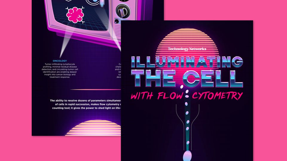
Flow cytometry continues to transform biological research by enabling rapid, high-dimensional analysis of millions of individual cells.
As researchers seek deeper insights into cellular heterogeneity, disease mechanisms, and therapeutic responses, advances such as spectral flow cytometry, mass cytometry, imaging flow cytometry, and AI-assisted analysis are expanding what is possible.
This infographic explores the core technologies behind flow cytometry and highlights the innovations driving new discoveries across immunology, oncology, and cell therapy.
Download this infographic to discover:
- How the four core systems of flow cytometry work together to generate single-cell data
- The latest innovations enabling higher-parameter analysis, improved signal resolution, and more reproducible workflows
- How advanced cytometry approaches are accelerating breakthroughs in immunology, oncology, and cell therapy research
Flow cytometry has become one of the most powerful tools in modern
biology, providing researchers with a range of abilities, from counting cells to
investigating their biology. By passing cells single-file through a focused laser
beam, it captures a multidimensional snapshot of each cell. This infographic
explores the power behind flow cytometry, as well as the innovations and data
analysis that are pushing the boundaries of what this technique can reveal.
THE CORE COMPONENTS OF FLOW CYTOMETRY
Flow cytometry's power lies in the precise coordination
of four key systems.
FLUIDICS
Hydrodynamic focusing
delivers cells single-file
through the interrogation
point, ensuring each cell is
analyzed individually
LASER EXCITATION
One or more lasers illuminate cells; modern
instruments can incorporate up to six lasers
spanning UV to near-infrared wavelengths
OPTICS AND DETECTORS
Bandpass filters and photomultiplier tubes
or avalanche photodiodes capture emitted
fluorescence and scatter signals
DATA ACQUISITION
Electronic systems convert optical signals
into digital data, with modern platforms
processing thousands of cells per second
RECENT INNOVATIONS IN CYTOMETRY
The field is undergoing a step-change in capability, driven by advances
across hardware, reagents, and computation.
Spectral flow cytometry
moves beyond
conventional filterbased
detection. Rather
than capturing discrete
emission peaks, spectral
instruments capture the
full emission spectrum of
each fluorochrome and
use unmixing algorithms
to separate overlapping
signals, enabling 40+
parameter panels from a
single sample.
Mass cytometry
replaces fluorescent
labels with stable
heavy metal isotopes
detected by timeof-
flight mass
spectrometry. With
minimal signal overlap,
panels exceeding 50
markers are routinely
achievable at the cost
of sample throughput.
Imaging flow cytometry
marries the statistical power of
conventional flow cytometry with
single-cell morphological imaging,
enabling spatial analysis of protein
localization, nuclear morphology,
and cell–cell interactions.
AI-assisted gating is replacing
manual population identification.
Machine learning tools now automate
and standardize gating strategies,
reducing operator variability and
enabling reproducible analysis of
high-dimensional datasets.
SHEDDING LIGHT ON BIOLOGY
These advances are accelerating discovery across a range of research areas.
IMMUNOLOGY
High-parameter immunophenotyping maps rare cell subsets
and activation states; spectral cytometry is redefining our
understanding of T cell heterogeneity and exhaustion.
ONCOLOGY
Tumor-infiltrating lymphocyte
profiling, minimal residual disease
detection, and circulating tumor cell
identification are enabling deeper
insight into cancer biology and
treatment response.
CELL THERAPY
CAR-T and NK cell product
characterization, potency testing,
and in vivo persistence monitoring
rely on multiparameter flow
cytometry at every stage of
development and manufacture.
The ability to resolve dozens of parameters simultaneously, across millions
of cells in rapid succession, makes flow cytometry more than just a
counting tool; it gives the power to shed light on life-changing biology.
By Steven Gibney, PhD | Designed by Erin Lemieux
SPONSORED BY