Virtual Reality Tool Helps Process Brain Scan Data
Complete the form below to unlock access to ALL audio articles.
In recent years, virtual reality technology has begun proving its worth outside the world of gaming, with applications in education, health care, military training and beyond. Now researchers at the Mark and Mary Stevens Neuroimaging and Informatics Institute at the Keck School of Medicine of USC are exploring a new frontier: VR as a tool for processing brain scan data.
“This project is particularly exciting because not much has been done with VR in the field of neuroscience,” said Arthur Toga, provost professor and director of the neuroimaging and informatics institute.
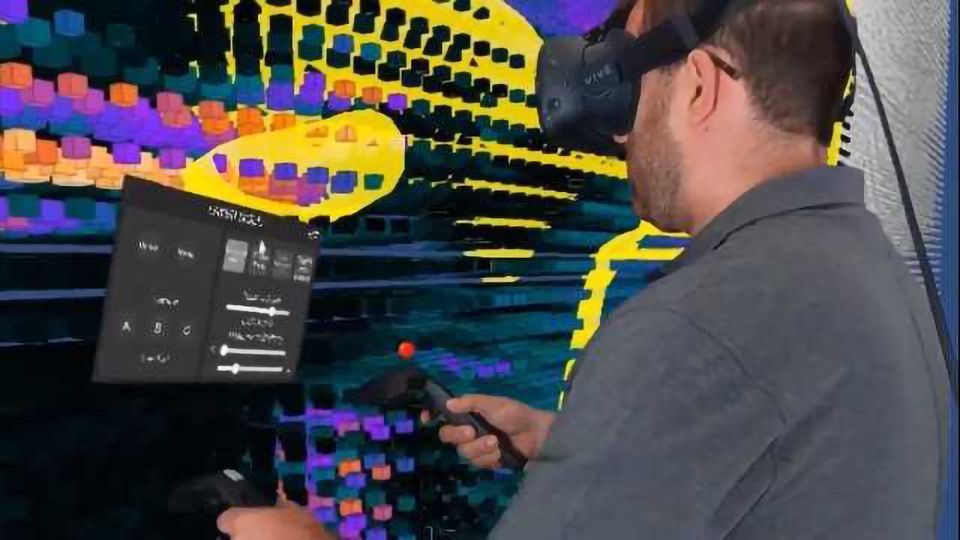
The new tool, Virtual Brain Segmenter (VBS), transforms a tedious step in the scientific process into an immersive experience. And a new study published in the Journal of Digital Imaging in July suggests that the tool significantly increases the efficiency of brain scan analysis.
Let the processing stage begin
After researchers collect MRI data from study participants, the processing stage begins. During automatic processing, one step, called segmentation, separates the brain into labeled regions to facilitate close examination of various structures. But the automation isn’t perfect, and researchers must manually correct errors before moving forward with analysis.
The time-consuming work requires a trained hand, so most labs hire undergraduate research assistants to correct segmentation errors. But with the vast amount of data being collected, studies still tend to hit a bottleneck during the processing stage. And many labs experience a high turnover of research assistants, who tire of the repetitive task, amounting to wasted time and resources spent training new workers.
“We wondered: How can we make this process more efficient and intuitive? Is there a way to correct segmentation errors that requires less training?” said Dominique Duncan, assistant professor of neurology at INI and lead author of the paper.
Experimental trial
With this in mind, Duncan, Toga and Assistant Professor Tyler Ard launched an experimental trial to test the efficacy of VR in brain segmentation. They teamed with RareFaction Interactive, which programmed VBS and assisted with its design, and secured a Vive VR headset from the consumer electronics company HTC. Duncan also received a research grant from the USC Postdoctoral Association to fund the trial.
The experiment tested 30 participants, all new to brain segmentation, on two tasks: using a MacBook to perform a minor error correction in the classic data cleanup program FreeSurfer, and completing the same task in VBS, armed with the HTC Vive headset and two controllers.

On average, participants finished the correction 68 seconds faster using VBS compared with FreeSurfer, a highly significant time savings considering the task rarely took more than three minutes to complete.
“Our main goal is to put this tool in the hands of researchers around the world,” Toga said. “We see that as a way to continue driving discovery in the field.”
This article has been republished from materials provided by the University of Southern California. Note: material may have been edited for length and content. For further information, please contact the cited source.
Reference: Duncan, D., Garner, R., Zrantchev, I., Ard, T., Newman, B., Saslow, A., … Toga, A. W. (2018). Using Virtual Reality to Improve Performance and User Experience in Manual Correction of MRI Segmentation Errors by Non-experts. Journal of Digital Imaging. https://doi.org/10.1007/s10278-018-0108-5

