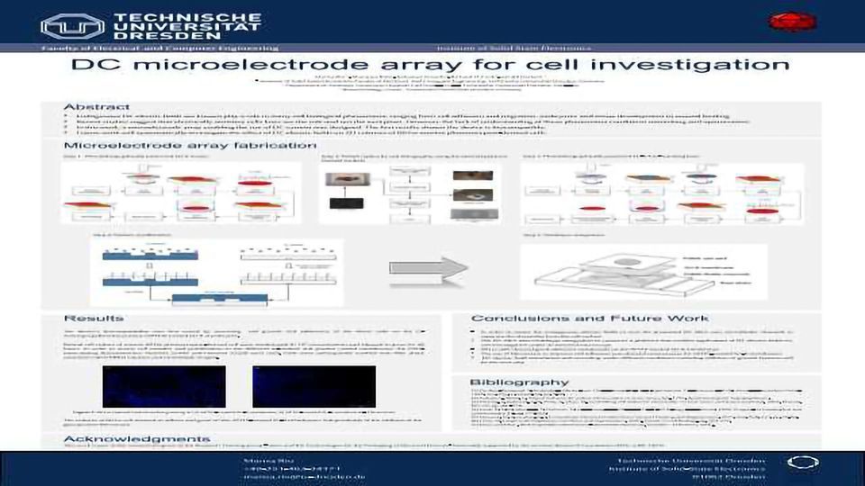
DC Microelectrode Array for Cell Investigation

The discovery that cells undergo electrotaxis, orient and migrate in a specific direction relatively to electric fields, dates for the late 19th century. Recent studies suggest that electrically sensitive cells lines are the rule and not the exception. The influence of endogenous DC electric fields is now shown to be present in many cell biological phenomena, ranging from cell adhesion and migration, embryonic and tissue development to wound healing.
In the present study, we report the steps towards the assembling of an electrically stimulated microfluidic biochip. In order to mimic the in vivo environment DC electric fields are used. This biochip intends to stimulate and record the response of cells to different DC electric fields. Its main feature involves the replacement of conventional metallic microelectrodes positioned directly beneath the cells surface by channels filled with conductive medium, therefore delaying Faradaic products to attain the cell surface. The biochip architecture is based on multilayer integration over a polycarbonate base plate containing Ag/AgCl electrodes.
The first results showed the biocompatibility of the microfluidic biochip and promise to render further understanding of the cellular interaction with DC electric fields.


