The Culture of Cell Culturing: Be Careful Not To Contaminate!

Complete the form below to unlock access to ALL audio articles.
In vitro cell culture is a vital aspect of basic and biomedical research. Cell culture can be readily and rapidly used to test a hypothesis and collect preliminary data before more time-consuming in vivo experiments. It is also a useful screening tool, for example for drug screens or genetic experiments, by virtue of ease of scaling and amenability to high throughput. The importance of cell culture to research cannot be understated. Nevertheless, it is frequently seen as an overture to more important or definitive in vivo experiments. Despite this impression, it is still critical to dedicate rigor and care to in vitro experiments, which will serve as the foundation to future experiments. Therefore, it is essential to apply the highest standards to cell culturing techniques. Researchers must consider a number of factors for in vitro experiments. Selecting the most appropriate cell line or primary culture for the project at hand is only the first step. Researchers must then be vigilant to prevent contaminating the culture, from the initial moments it is plated and all throughout consecutive passages.
Culture contaminants
One source of culture contamination is the inadvertent introduction of another cell line due to poor experimental technique or gross human errors. “This is a real concern in cancer research,” explains Vivekan Nand Yadav, PhD, who is a research investigator in pediatric gliomas in the Department of Pediatrics-Hematology/Oncology at the University of Michigan. “There are numerous instances of these so called problematic cell lines, that became contaminated with another cell line, which outgrows the original one.” A prominent and well-known cause of problematic cell lines is the HeLa cell line, which was derived from an aggressive cervical adenocarcinoma. HeLa cells proliferate so efficiently that they can overrun the contaminated culture. “It is important to keep track of these problematic cell lines as they plague biomedical research and confound study results,” says Yadav. “Databases are a good resource, such as Cellosaurus, which hosts a page dedicated to problematic cell lines that are either contaminated or misidentified. Another good practice is to report Research Resource Identifiers (RRIDs) for cell lines used in any publication. RRIDs are unique identifiers for biological resources, including cell lines, and was developed to boost reproducibility and transparency in biomedical research.”
Cell cultures can also become contaminated with varieties of microorganisms, such as bacteria or fungi. “You’ll know immediately when you have a bacterial contamination in your cultures,” says Yadav. “They’re generally very visible and odorous and grow so efficiently that they turn your culture media cloudy. Fungal contaminations are identifiable by microscopic examination. Yeasts and filamentous fungi appear as bright ovoids or fibers, respectively. A really bad mold infestation will also be visible to the eye. For both bacterial and fungal issues, you have to discard the cultures at this point, appropriately through biohazard, clean the culture room, check stock solutions and throw out any with a contamination, and start over,” advises Yadav.
He continues: “The more insidious problem is mycoplasma, which is a widespread issue that is far harder to detect.”. Mycoplasma are the smallest self-replicating microorganism, a type of bacteria devoid of a cell wall that allows them to adopt many shapes, which camouflages them in culture. “Mycoplasma hijack nutrients from cultured cells and may influence their growth, gene expression and cellular status. This is a problem for research. For example, in my projects, I investigate mechanisms of tumor reprogramming and drug susceptibility in adult and pediatric gliomas. These are cellular properties that mycoplasma can influence, which would render data interpretation impossible. Therefore, I always ensure my cultures are free from of contaminating mycoplasma before performing my experiments.”
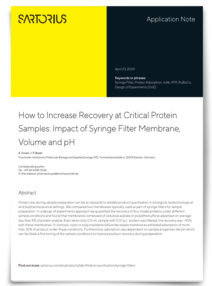
How To Increase Recovery at Critical Protein Samples: Impact of Syringe Filter Membrane, Volume and pH

A common occurrence in product quantitation is the loss of proteins, which can ultimately lead to distorted results and inaccurate data. One approach to counter analyte loss is the use of membrane filters however, to be used accurately the membrane must consist of the appropriate pH, surface charge, and handling steps relevant to the proteins under analysis. Download this app note to discover the different membranes used in protein analysis and how the materials of filter membranes differ in protein adsorption and which are most effective.
Download App NoteSponsored Content
Readily recognizing problems
With the quality of research at stake, vigilance is essential. “You need to invest time to validate your cell line before embarking on a project,” advises Zsolt G. Venkei, PhD, a research scientist at the Whitehead Institute for Biomedical Research, Cambridge US. “This is especially critical when using a cell line that is new to the lab. This process of cell line authentication can be performed in many ways. The easiest method, which should be integrated into your cell culture routine, is quick visual microscopic inspection for morphology. This is a crude approach, because cell line morphologies are not unique; however, a shift in shape can alert to a contamination problem. I’d say the best method is short tandem repeat profiling, a PCR-based assay that matches a cell’s variable DNA microsatellite profile to a database. But other genomic approaches are also useful, such as karyotyping and copy number variation such as by G-banding and fluorescence in situ hybridization (FISH).”
Venkei utilizes Drosophila as a research model and has worked in insect cell culture. Cell line provenance, the species and tissue origin of the cell line, is another important aspect of the authentication process. “When you are conducting research, you are trying to answer questions in a specific model organism or cell line. One way to test provenance, is to perform an isoenzyme analysis to validate species origin and immunocytochemistry for biomarkers of tissue origin,” he explains. “This way, you have validated the authenticity of your cell line, and checked that it maintains the characteristics you need to address your research question.”
“Past the authentication process, one must now ensure no microorganisms contaminate your culture as you passage it and conduct experiments,” says Venkei. “Microorganism contamination ranges from the obvious bacterial and fungal, which are visible, either by eye or microscope, to mycoplasma, which do not leave readily discernable visual signs. Mycoplasma are generally difficult to detect by microscopy, and are usually detected by molecular techniques, such as PCR and FISH, which should follow a routine schedule, since there are limited visual signs to alert to a possible contamination. Finally, viruses are bit trickier. Some viruses induce a cytopathic effect, a morphological change, that is visually detectable. Others may simply integrate into the host cell DNA without affecting cell morphology, in which case a molecular test such as PCR is required.”
Best Practices for Cell Culture Confluency Calculation With Incellis

Cell confluency is an important technique to be applied with accuracy, as it is a costly process which requires a maximum efficiency of transfection from the first attempt. It is a process in which a sample is analyzed by the observation of space occupied by cells – this can be amplified via utilization of hemocytometers and dyes such as Trypan blue, but these additional steps require more work. Bertin Technologies have developed a new method that allows for easier use of cell confluency. Download this whitepaper to discover how the “scratch” assay method measures cell migration in vitro and how a novel cell imaging system can provide cost-effective, high specificity results.
View WhitepaperSponsored Content
Preventing problems preemptively
Almost all contamination issues can be minimized by implementing appropriate safeguards. “The first measures you can take are in the culture itself,” explains Venkei. “Make sure you use sterile plasticware or sterilized glassware, include antibiotics and antimycotics in your culture media, and check the culture daily. Always be especially vigilant with new cell lines acquired to the lab. Check them for mycoplasma, because once a contamination sets in, it is hard to eradicate,” Venkei cautions. The next measures are to ensure a clean work environment. Always work in a laminar flow hood; follow the instructions for sash level and do not obstruct the flow by cluttering the hood and covering the vents. Make sure the laminar flow hood filter is routinely serviced and replaced. Venkei elaborates on some of the key safeguarding measures: “Decontaminate the hood by UV light, and all equipment you take into it with 70% ethanol. Make sure the incubator you store your cultures in is routinely cleaned. When you work, follow good pipetting technique to prevent aerosolization and minimize spread. Wear a lab coat and gloves, as much to protect yourself as your culture.”
Generally, preventing contamination is more straightforward than eradicating a persistent contamination, which requires more extreme measures, such as discarding cultures and opened media, antibiotics/antimycotics, and sera bottles. Precious or irreplaceable cultures can be salvaged, but require special care. “It’s a lot of steps, a lot of measures, a lot of work. But all of it necessary to maintain quality cell cultures, free from contamination, and to ensure the integrity of your research,” concludes Venkei.


