Going Label-Free: The Future of Metabolomics Imaging

Complete the form below to unlock access to ALL audio articles.
The study of brain tumors using real-time imaging of the brain in live disease models is critical for elucidating disease mechanisms. It is also essential for the discovery of treatment targets for the development of novel intervention strategies. Such research requires full characterization of healthy and disease phenotypes using a multi-omics analysis, incorporating genomics, transcriptomics, proteomics and metabolomics.
Metabolomics is the scientific study of biochemical processes involving metabolites. These processes are highly dynamic and, as such, can pose a challenge for scientists looking to observe and measure these changes. Despite being one of the biggest emerging areas of the life sciences and the focus of intense investment in research, imaging of the metabolome and the discovery of metabolomic biomarkers indicating pathology remains a technological challenge.
Fluorescent dyes can be effective for defining structures, but their use in the laboratory and clinic is limited, particularly for live imaging. Obtaining meaningful fluorescence microscopy data from live cells is challenging as photodamage needs to be minimized while retaining a useful signal-to-noise ratio. A suitable environment also needs to be maintained for cells and tissues to replicate physiological cell dynamics. There is therefore a growing trend towards the use of label-free multiphoton imaging.
Why we need tunable detection
The SP8 Deep In Vivo Explorer (DIVE) microscopy system is the first multiphoton microscope with spectrally tunable non-descanned detection (4TUNE). Multiphoton (MP) microscopy, often also referred to as two-photon microscopy, uses infrared (IR) light. It has the great advantage of being able to penetrate deep into the sample – up to 1 mm and beyond (depending on the nature of the tissue), being less phototoxic and able to excite not only classical fluorescent labels but also intrinsic fluorescence such as nicotinamide adenine dinucleotide (NADH). Furthermore, the intrinsic confocality of a MP excitation process offers improved z-sectioning. These benefits make MP microscopy the preferred tool for deep in vivo imaging.
The 4TUNE capability of SP8 DIVE offers researchers the flexibility of visualizing samples in multicolour (Figure 1). Moreover, the introduction of a variable beam expander (VBE) enables the user to choose between best resolution and best depth in order to achieve the highest resolution and penetration depths of more than 1 mm, making this an ideal tool for visualizing deep into thick, live samples (Figure 2).
1583255313548.jpg) Figure 1: 4Tune enables new dye combinations: Confetti mouse small intestine. Cell lineage tracer. Cyan: CFP, green: GFP, yellow: YFP, red: RFP. Colon cancer research. Credit: Prof. Jasco van Rheenen, Netherlands Cancer Institute, Amsterdam (the Netherlands).3
Figure 1: 4Tune enables new dye combinations: Confetti mouse small intestine. Cell lineage tracer. Cyan: CFP, green: GFP, yellow: YFP, red: RFP. Colon cancer research. Credit: Prof. Jasco van Rheenen, Netherlands Cancer Institute, Amsterdam (the Netherlands).3 Figure 2: Mouse brain, transgenic and transiently labeled. Different types of nerve cells are shown in green (microglia, GFP), yellow (neurons, YFP), blue (astrocytes, sulforhodamine) and the blood system is shown in red (Alexa680-dextran). Credit: Kevin Keppler, Light Microscope Facility, DZNE Bonn (Germany).3
Dr Thomas Mathivet, from the Cardiovascular Institute in Paris, France, uses in vivo imaging of the cardiovascular system in the brain to investigate ways of curing blood vessel damage in the context of treating diseases such as strokes and brain tumors like gliomas.1 He says, “In my previous lab, using another solution which required changing filters, we were restricted. With all the new models we are working on, which need five or six different channels for visualization at the same time, using filters was no longer viable. We could not image all this information together, which was really a pity. The DIVE system with the 4TUNE really changed what we could see. Basically, we can narrow the windows to 10 to 20 nm with those tunable detectors which are fantastic to image, for example, the Brainbow mouse, where you will have a green fluorescent protein, yellow fluorescent protein, red fluorescent protein and cyan fluorescent protein.”
FLIM for the win
MP imaging offers not only spectral and intensity-based information on the sample, but, when combined with fluorescence lifetime microscopy (FLIM), additional contrast can be obtained. FLIM is an imaging technique that produces an image based on the time that fluorophores spend in the excited state. In FLIM, it is the lifetime of the fluorophore signal, rather than its intensity, that is used to determine the contrast of each pixel and calculate and represent the digital image.
Fluorescence lifetime depends on the microenvironment in which the fluorophore is and more importantly, it is independent of the concentration of the fluorophore. Therefore, fluorescence lifetime is the method of choice for functional imaging, as it can be utilized for making measurements in chemical sensing, such as of the pH of the local molecular environment. FLIM can be applied in combination with multiphoton imaging to study metabolic processes or to follow oxidative stress without the need for labelling by analysing the intrinsic fluorescence of NADH, vitamins and other key biological cofactors (Figure 3).4
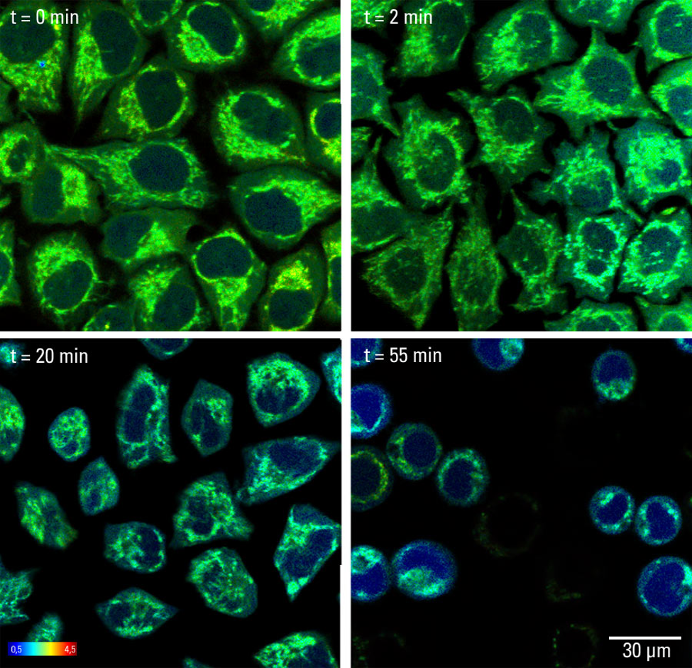 Autofluorescence in mammalian cells at non-physiological conditions (pH 8.5). The signal correlates with changes in the NAD/NADH endogenous pool. The development of oxidative stress reads out as decrease of fluorescence lifetime over time. Original image size: 512 x 512 pixels. Color bar scale (lifetime): ns. Two-photon image acquisition performed with SP8 DIVE and SP8 FALCON. Credit: Leica.
 The SP8 FALCON (FAst Lifetime CONtrast) system is a fully integrated Leica FLIM solution that enables FLIM at high speeds. With this instrument, complex FLIM experiments to study and track highly dynamic cellular physiology in living cells are now accessible for life scientists on a daily basis.
Future reveals of fluorescent microscopy
The continued advances in fluorescent microscopy promise to enable us to see and understand more about the functioning of cells and tissues in real-time and in-depth. With such imaging systems becoming more widely applied in the life sciences, it is anticipated that research using fluorescent microscopy will continue to reveal novel functional information, with the techniques becoming a standard tool routinely used to investigate metabolomic processes and cellular microenvironments. This will ultimately provide us with the knowledge and understanding we need to improve human health and create new treatments and innovative cures for human disease.
References:
1. Leica Microsystems. Chat with Dr. Thomas Mathivet about In Vivo Cardiovascular Imaging of the Brain. https://www.youtube.com/watch?v=uHMQURw1ges (accessed December 2019).
2. Alvarez LAJ, Widzgowski B, Ossato G, van den Broek G, Jalink K, Kuschel L, Roberti MJ, Hecht F. Application Note: SP8 FALCON: a novel concept in fluorescence lifetime imaging enabling video-rate confocal FLIM. Nature Methods; 2019. https://www.nature.com/magazine-assets/d42473-019-00261-x/d42473-019-00261-x.pdf (accessed November 2019).
3. Leica Microsystems. SP8 DIVE – Deep In Vivo Explorer. https://downloads.leica-microsystems.com/Leica%20SP8%20DIVE/Brochures/SP8_DIVE_Brochure_Web_EN.pdf (accessed December 2019).
4. Stringari C, Cinquin A, Cinquin O, Digman MA, Donovan PJ, Gratton E. Phasor approach to fluorescence lifetime microscopy distinguishes different metabolic states of germ cells in a live tissue. Proc Natl Acad Sci U S A. 2011;108(33):13582–13587. doi:10.1073/pnas.1108161108.

