Dysfunctional Mitochondria Initiate Parkinson’s Onset
Research indicates that mitochondrial dysfunction triggers a cascade of cellular changes that drive Parkinson’s disease.
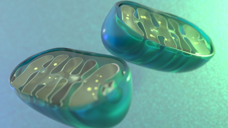
Although researchers have known for decades that mitochondria malfunction in Parkinson’s disease, it’s unclear whether mitochondrial dysfunction causes or is caused by Parkinson’s.
New research from the Gladstone Institutes has demonstrated that failing mitochondria can initiate Parkinson’s onset.
Modeling Parkinson’s disease
Parkinson’s disease is the second most common neurodegenerative disorder, with the number of people with Parkinson’s worldwide expected to double to 25 million by 2050. It is characterized by movement-related symptoms such as tremor and stiffness, but symptoms can also include memory problems and issues with sleep. Symptoms are caused by the brain losing its ability to produce dopamine, which is involved in coordinating movement and regulating mood, memory and motivation.
Progress in understanding the pathology of Parkinson’s disease has been slow, in part due to the limitations of animal models used in research. The many forms and subtypes of Parkinson’s disease, and the different genetic and environmental drivers, makes the creation of accurate models challenging.
When some mitochondrial mutations that are linked to Parkinson’s disease in humans are represented in mouse models, the models can fail to develop the key features of the common, sporadic form of Parkinson’s that develops later in life.
The new study created and used a unique mouse model that carries a mutation in a mitochondrial protein called CHCHD2. The mice exhibit symptoms of a rare, inherited form of Parkinson’s that is indistinguishable from the sporadic form.
With this model, “we were able to watch, step by step, how mitochondria start to fail and how this process eventually leads to the accumulation of alpha-synuclein, the protein that builds up in pathological alterations in the brain called Lewy bodies in nearly all Parkinson’s patients,” said Dr. Kohei Kano, a postdoctoral fellow at the Gladstone Institutes and a co-first author of the study.
A cascade of steps that trigger cellular changes
The researchers performed a comprehensive phenotypic analysis of the CHCHD2 mutant mice, and compared them to mice without the mutation. They compared lifespan, motor function, dopamine receptor function and mitochondrial ultrastructure.
The mutated CHCHD2 protein accumulated in mitochondria, causing them to swell and become distorted, disrupting protein-protein interactions and resulting in dysfunction.
The impairment of mitochondria shifted cellular metabolism, promoting a less efficient energy production pathway that resulted in the build-up of reactive oxygen species. The CHCHD2 mutation appeared to interfere with proteins that usually remove these destructive molecules, further increasing oxidative stress within the cells.
Alpha-synuclein began to accumulate after the levels of reactive oxygen species rose.
“This order of events is consistent with our hypothesis that oxidative stress is causing the alpha-synuclein to aggregate,” said Dr. Szu-Chi Liao, co-first author of the paper.
The buildup and aggregation of alpha-synuclein plays a key role in the progressive death of neurons in Parkinson’s disease.
“This mouse model provides some of the most compelling evidence to date for how mitochondrial dysfunction can cause typical late-onset Parkinson’s disease,” Dr. Ken Nakamura, who led the study, said.


Navigating Common Pitfalls in Gene Therapy Development
This webinar will explore lessons learned from previous development programs, including problems encountered by early researchers and the approaches used to address them.View Webinar
The researchers also examined post-mortem brain tissue from people with sporadic Parkinson’s to confirm their findings. In the human patients, the mutated CHCHD2 protein accumulated in dopamine-producing neurons during the early stages of alpha-synuclein aggregation.
A blueprint for Parkinson’s
“This work is a blueprint for how a mitochondrial protein can be disrupted and actually cause Parkinson’s disease,” Nakamura said. “There could be other triggers that set off this same sequence of events involving mitochondrial damage, energy problems, the accumulation of reactive oxygen species and finally, abnormal accumulation of additional proteins.”
The researchers plan to conduct additional studies to understand how CHCHD2 influences oxidative stress and whether it contributes to the development of sporadic Parkinson’s.
Understanding the link between mutated CHCHD2, mitochondrial dysfunction and the onset of Parkinson’s disease may also point to new drugs, such as those that block reactive oxygen species, with the hope of stopping the disease pathway.
Reference: Liao SC, Kano K, Phanse S, et al. CHCHD2 mutant mice link mitochondrial deficits to PD pathophysiology. Sci Adv. 2025;11(46):eadu0726. doi: 10.1126/sciadv.adu0726
This article is a rework of a press release issued by the Gladstone Institutes. Material has been edited for length and content.
 Add Technology Networks as a preferred Google source to see more of our trusted coverage.