NutriStem® hPSC XF Medium A Superior Xeno-free, Serum-free Culture Medium for hES and hiPS Cells
Expanding the culture of excellence
NutriStem® hPSC XF Medium is a defined, xeno-free, serum-free medium designed to support the growth and expansion of human induced pluripotent stem (hiPS) and human embryonic stem (hES) cells in a feeder-free environment. NutriStem® hPSC XF Medium offers the ability to culture human pluripotent cells without the need for high levels of bFGF and other stimulatory growth factors or cytokines. The low-protein formulation contains only the most essential components required for maintenance of hES and hiPS cells, providing a simplified medium and maintaining the cells’ full differentiation potential. The defined, xeno-free formulation of NutriStem® hPSC XF Medium provides consistent media performance and predictable cellular behavior, as well as increased reproducibility in long-term culture (over 50 passages). In addition, cells cultured in NutriStem® hPSC XF Medium show superior attachment and proliferation rates, making this medium ideal for high-throughput screening applications.

Figure 1: NutriStem® hPSC XF Medium enables excellent proliferation of undifferentiated hES and hiPS cells. Proliferation of H1 hES cells cultured in Matrigel-coated 96-well plates in NutriStem® hPSC XF Medium and the leading competing medium for feeder-free culture. Medium was changed and proliferation was assessed every 24 hours in culture
Normal cell morphology and functional assesment of pluripotency
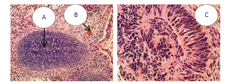
The formation of compact colonies of cells with a high nucleus-to-cytoplasm ratio, prominent nucleoli, and distinct colony borders are characteristic morphology traits of healthy undifferentiated hES and hiPS cells, and can be observed through a phase-contrast microscope (Figure 2). Human pluripotent stem cells hold the potential to differentiate into cell types of all three germ layers, i.e., endoderm, mesoderm, and ectoderm. This differentiation potential is assessed by the spontaneous differentiation within embryoid bodies cultured in vitro (Figure 3) and teratomas formed in vivo (Figure 4).

Figure 2: Normal Colony Morphology.H1 hES cells (panel A) and ACS-1014 hiPS cells (panel B) cultured in NutriStem® hPSC XF Medium on Matrigel-coated plates displaycolony morphologies typical of normal feeder-free hES and hiPS cell cultures, including a uniform colony of tightly compacted cells and distinct colony edges.
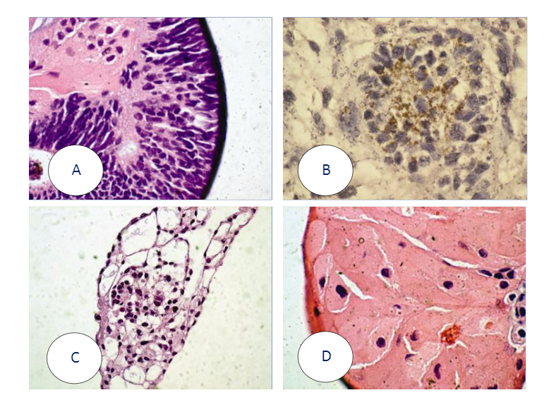
Figure 3: Embryoid Body Formation. Embryoid bodies (EBs) were generated from H9.2 hES cells cultured for 16 passages in NutriStem® hPSC XF Medium on Matrigel matrix as an evaluation of pluripotency. The pluripotent H9.2 cells were suspended in serum-supplemented medium, where they spontaneously formed EBs containing cells of embryonic germ layers. The following cell types were identified by examination of the histological sections of14-day-old EBs stained with H&E: (A) neural rosette (ectoderm), (B) neural rosette stained with Tubulin, (C) primitive blood vessels (mesoderm), and (D) megakaryocytes (mesoderm).