Tandem Mass Spectrometry (MS/MS) Explained
This comprehensive overview details the mechanisms of tandem MS, and compares HCD vs CID fragmentation.
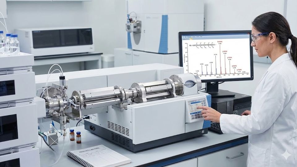
Tandem mass spectrometry serves as the cornerstone of modern protein sequencing and identification workflows in clinical and research laboratories. Implementing MS/MS in proteomics allows laboratory professionals to determine the primary amino acid sequence of peptides with high specificity by isolating and fragmenting precursor ions. This technique provides the structural information necessary to characterize complex biological samples, identify post-translational modifications (PTMs), and validate potential biomarkers for disease states. While single-stage mass spectrometry can measure the mass of an intact analyte, it cannot definitively establish sequence identity without the fragmentation patterns generated by tandem stages. Understanding the distinct physical mechanisms behind various fragmentation modes ensures the selection of the most appropriate method. This knowledge is also crucial for evolving acquisition strategies like Data-Independent Acquisition (DIA). This article explores the operational principles of tandem MS, compares dissociation techniques, and discusses best practices for data interpretation in a high-throughput environment.
Fundamentals of MS/MS in proteomics workflows
Effective protein characterization depends on the precise isolation and fragmentation of peptide ions within the mass spectrometer, typically coupled with liquid chromatography (LC-MS/MS). In a typical "bottom-up" proteomics experiment, proteins undergo enzymatic digestion—usually with trypsin—to produce peptides. These peptides are separated by hydrophobicity on a reversed-phase column before entering the mass spectrometer via electrospray ionization (ESI). The instrument first measures the mass-to-charge ratio (m/z) of the intact peptide ions (MS1). To gain structural information, the instrument must then select specific precursor ions for fragmentation (MS2). This process involves adding energy to the ions, causing them to break apart into smaller fragment ions. The resulting tandem mass spectrum contains a pattern of peaks representing these fragments, which bioinformatics software uses to reconstruct the peptide sequence.
The integration of MS/MS in proteomics requires sophisticated instrumentation capable of multiple stages of mass analysis. Common configurations include triple quadrupole (QqQ), quadrupole-time-of-flight (Q-TOF), and orbital ion trap hybrid instruments. Triple quadrupoles excel at targeted quantification using selected reaction monitoring (SRM) or multiple reaction monitoring (MRM), offering high sensitivity for known targets. Conversely, Q-TOF and orbital trap instruments provide the high resolution and mass accuracy needed for discovery proteomics. A critical evolution in this workflow is the shift from Data-Dependent Acquisition (DDA) to Data-Independent Acquisition (DIA). In DIA, the instrument fragments all ions within wide isolation windows. This creates complex chimeric spectra that require advanced deconvolution but provide a permanent digital archive. Laboratory professionals must calibrate these systems rigorously to ensure that the selection of precursor ions and the subsequent fragmentation energy yield informative spectra.
Key instrumentation components
Quadrupole mass filter: Isolates specific precursor ions based on their m/z values. It acts as a gatekeeper that allows only ions of interest (or ranges of ions in DIA) to pass.
Collision cell: Facilitates the interaction between precursor ions and inert gas molecules (typically nitrogen or argon) or reagent ions to induce fragmentation.
Mass analyzer: Detects and measures the m/z of the resulting fragment ions with high precision, with orbital traps and TOFs offering resolving power sufficient to distinguish isotopic envelopes.
Comparing fragmentation methods including HCD vs CID
Selecting the appropriate dissociation technique determines the type of fragment ions produced and the resulting sequence coverage. The internal energy transfer during the collision process dictates where the peptide backbone breaks. Two of the most prevalent methods in beam-type and trap-type instruments are Collision-Induced Dissociation (CID) and Higher-energy Collisional Dissociation (HCD). Both methods rely on collisions with neutral gas molecules. However, they differ significantly in how energy is applied, the activation timeframe, and how fragments are detected.
Collision-induced dissociation (CID)
CID remains a standard fragmentation method, particularly in legacy ion trap instruments and some tribrid workflows. In this process, precursor ions accelerate and collide with an inert gas such as helium. This collision converts kinetic energy into internal vibrational energy, causing the peptide bond to break primarily at the amide linkage. This cleavage generates b-type (N-terminal) and y-type (C-terminal) ions. CID is highly effective for general peptide sequencing but faces limitations when analyzing low-mass fragments. In resonance-excitation ion traps, the "low-mass cutoff" (typically one-third of the precursor m/z) prevents the detection of diagnostic ions, such as those for isobaric tags or specific immonium ions. Consequently, while CID produces rich spectra for sequence confirmation, it is often unsuitable for quantification workflows that rely on low-m/z reporter ions.
Higher-energy collisional dissociation (HCD)
HCD overcomes many limitations of traditional trap-based CID and has become the gold standard for high-resolution discovery proteomics. Originally developed for orbital ion trap instruments, HCD occurs in a dedicated multipole collision cell where ions undergo beam-type fragmentation, similar to that in a triple quadrupole. This method allows for the detection of ions across the entire mass range, effectively eliminating the low-mass cutoff issue associated with resonance CID. Consequently, HCD proves superior for detecting reporter ions from isobaric labeling experiments. It also excels at detecting diagnostic ions for modifications like phosphorylation (e.g., the tyrosine immonium ion). The resulting spectra typically exhibit high mass accuracy and a rich ion series, enhancing the confidence of MS/MS in proteomics identification. Furthermore, HCD fragmentation energies can be normalized, allowing for more reproducible fragmentation across different precursors and instrument platforms.
Table 1. A comparison of common fragmentation techniques.
| Feature | Collision-induced dissociation (CID) | Higher-energy collisional dissociation (HCD) |
| Primary mechanism | Resonant excitation with gas collisions (slow heating) | Beam-type collisions in a dedicated cell (fast heating) |
| Ion types | Primarily b- and y-ions | b- and y-ions, often with immonium ions |
| Low-mass cutoff | Yes (approx. 1/3 precursor m/z) | No (full mass range detection) |
| Primary use case | Routine peptide identification, spectral library building | Isobaric tagging, phosphoproteomics, de novo sequencing |
| Instrumentation | Ion Traps (Linear, 3D), Tribrids | Orbitrap Hybrids, Q-TOFs |
The role of electron transfer dissociation (ETD)
Electron transfer dissociation provides an alternative fragmentation pathway essential for analyzing labile post-translational modifications (PTMs) and large peptides. CID and HCD use vibrational energy that often "knocks off" fragile modifications (such as phosphate or glycan groups) before the peptide backbone breaks. Unlike these methods, ETD uses ion-ion chemistry. The process involves transferring an electron from a radical anion to the multi-charged peptide cation. This transfer induces radical-driven cleavage of the N-Cα bond, producing c-type and z-type ions rather than the b/y ions seen in collisional methods.
This "soft" fragmentation technique preserves labile side-chain modifications. When analyzing O-GlcNAcylation or phosphorylation, standard vibrational methods often strip the modification, leaving the peptide sequence ambiguous regarding the site of attachment. ETD leaves the modification attached to the specific amino acid residue, allowing for precise site localization. Consequently, laboratories focusing on PTM analysis often employ a decision-tree method. The instrument switches between HCD and ETD based on precursor charge state and m/z, as ETD favors higher charge states.
Advanced hybrid methods combine both electron transfer and collisional activation in a single spectrum. This dual approach generates c/z ions (preserving PTMs) and b/y ions (providing backbone sequence) simultaneously. This offers the most comprehensive characterization available for complex glycoproteins or large middle-down peptides. Organizations like the Human Proteome Organization (HUPO) and the National Cancer Institute's Clinical Proteomic Tumor Analysis Consortium (CPTAC) emphasize the importance of using such complementary fragmentation methods. By combining HCD and ETD spectra, laboratories can achieve higher confidence in characterizing modified peptides that might otherwise remain unidentified.
Spectral interpretation and data analysis strategies
Accurate peptide identification depends on the systematic correlation of experimental mass spectra with theoretical databases. Once the mass spectrometer generates the tandem MS data, the burden shifts to bioinformatics pipelines. The software algorithms compare the observed fragment ions against theoretical spectra generated from a reference proteome (e.g., UniProt). Essential factors in this process include the mass accuracy of the precursor and fragments, the signal-to-noise ratio, and the complexity of the sample matrix.
Successful spectral interpretation requires a solid understanding of ion series. In a typical tryptic peptide fragmented by HCD, y-ions usually dominate the high-mass region of the spectrum, while b-ions appear in the lower range. Proline residues often produce intense fragmentation N-terminal to the proline, creating a prominent gap or peak. Conversely, interpreting ETD spectra requires looking for c- and z-ion series. Analysts must also account for neutral losses (e.g., loss of water or ammonia) which can complicate the spectrum. For Data-Independent Acquisition (DIA) data, the analysis is distinct. Rather than one-to-one spectrum matching, the software must "deconvolute" complex mixed spectra. This often uses spectral libraries or deep learning-predicted libraries to extract quantitative data.
Validating these results involves statistical scoring, typically using a Target-Decoy approach to estimate the False Discovery Rate (FDR). Laboratories generally accept an FDR of 1% at the peptide and protein levels. However, manual inspection of spectra remains a valuable skill for validating borderline cases, novel modifications, or de novo sequencing results where no database exists. During manual review, analysts verify that the major peaks in the spectrum correspond to the assigned sequence and that the intensity distribution matches expected fragmentation behavior. This rigorous verification ensures that the MS/MS in proteomics data supports the biological conclusions drawn from the study.
Quality control and system suitability testing
Maintaining data integrity in MS/MS in proteomics requires rigorous quality control (QC) and system suitability testing (SST). Before analyzing valuable biological samples, the laboratory must analyze a standardized QC sample, such as a HeLa cell lysate digest or a mixture of bovine serum albumin (BSA) peptides. This step verifies that the liquid chromatography (LC) system delivers reproducible retention times and that the mass spectrometer achieves the expected sensitivity and mass accuracy. Monitoring parameters such as the number of peptide spectral matches (PSMs), peak width (FWHM), and injection time stability helps detect instrument drift or contamination early.
Modern laboratories often utilize automated QC software to plot these metrics longitudinally. A sudden drop in identified peptides might indicate a dirty ion transfer tube or a failing column, while a shift in mass accuracy suggests a need for recalibration. Establishing a longitudinal QC protocol allows the laboratory to track performance over time, ensuring that variations in data result from biological differences rather than instrumental instability. Regulatory bodies like the FDA encourage the documentation of SST performance as part of biomarker qualification guidelines, underscoring its necessity in robust analytical workflows. Without these controls, the high sensitivity of modern MS/MS instruments becomes a liability, as they will readily detect contaminants or generate noise that mimics signal.
Future outlook for MS/MS in proteomics
Tandem mass spectrometry remains the definitive tool for protein sequencing and characterization in modern life sciences. By mastering the nuances of MS/MS in proteomics, including the selection of HCD versus CID or the application of ETD, laboratory professionals can extract maximum information from complex samples. As instrumentation continues to evolve toward higher speed and sensitivity, the integration of multiple fragmentation modes, hybrid acquisition strategies like DIA, and advanced bioinformatics will further refine the accuracy of peptide identification. Adhering to strict quality control standards and understanding the underlying physics of spectral generation ensures that proteomic data remains reliable and reproducible.
This content includes text that has been created with the assistance of generative AI and has undergone editorial review before publishing. Technology Networks’ AI policy can be found here.
 Add Technology Networks as a preferred Google source to see more of our trusted coverage.