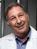
An Interview with Professor Fred Kramer

Want to listen to this article for FREE?
Complete the form below to unlock access to ALL audio articles.
Read time: 4 minutes
 Select Biosciences South East Asia recently interviewed Professor Fred Kramer from Rutgers University.
Professor Kramer is a member of the Department of Microbiology, Biochemistry, and Molecular Genetics at Rutgers University. After receiving a doctorate from the Rockefeller University, he was a member of the faculty of Columbia University for 17 years, and then moved his laboratory to the Public Health Research Institute, where he has carried out research with his colleagues for the past 29 years.
Select Bio SEA: After your undergraduate studies in Zoology, you completed a Ph.D. with Vincent Allfrey at The Rockefeller University. How did this experience influence your future research interests?
Professor Kramer: I set out to become a developmental biologist, and I tackled the problem of how maternal messenger RNAs are stored in frog oocytes for use after fertilization. This led to the development of in vitro assays that assessed the amount of stored messenger RNAs. As a postdoctoral fellow at Columbia, I continued working on in vitro assays that probed the mechanism of exponential replication of RNA by bacteriophage replicases, and that led to in vitro selection experiments in which RNA populations evolved in response to the presence of inhibitors of replication. Before long, the nucleic acid molecules themselves became my “experimental animals,” and the generation of new nucleic acid molecules whose structure and function could be designed for useful purposes became my passion.
Select Bio SEA: Your lab has a long history of exploring nucleic acid structure. Can you describe some of the projects you have worked on over the years and some of the experimental techniques you have developed?
Professor Kramer: Here are some highlights: (1) in 1989 we introduced the use of intercalating dyes to follow the exponential amplification of nucleic acids in real time, and we noted that the time that it takes to synthesis a particular amount of nucleic acid is inversely proportional to the logarithm of the number of template nucleic acids originally present in an assay (this approach is the basis of real-time PCR assays); (2) in parallel with Fred Sanger’s laboratory, we developed the chain-terminating method of nucleic acid sequence analysis, and we introduced the use of inosine as a means of preventing the formation of structures that obscure the sequencing results during electrophoresis; (3) we developed extremely sensitive clinical assays based on bifunctional recombinant RNAs that serve as probes, and that are then exponentially amplified by incubation with Q-beta replicase to indicate the amount of target present; (4) we invented molecular beacons, which are fluorescently labelled hybridization probes that are dark when free in solution, but that become brightly fluorescent in a preselected colour when they hybridize to target amplicons in PCR assays; and (5) we introduced the use non-FRET label pairs that interact by contact quenching, thereby enabling many different probes to be used simultaneously in highly multiplex clinical diagnostic assays.
Select Bio SEA: Your current research involves quantitation of extremely rare mutations associated with cancer. Can you tell us more about SuperSelective PCR Primers and how these are used in your research?
Professor Kramer: SuperSelective PCR primers are designed to initiate the amplification of a mutant sequence, while ignoring the corresponding wild-type sequence, even if the difference between the mutant and the wild type is only a single-nucleotide polymorphism. Two principles underlie the extraordinary selectivity and specificity of these primers: (1) thermodynamically, due to the extremely short hybrids that they form, the perfectly complementary hybrids formed by the primer with the mutant target sequence are considerably more abundant than the mismatched hybrids formed by the primer with the wild-type target sequence; and (2) because these short hybrids only last for less than one second before they fall apart, and because the mismatched wild-type hybrids persist for far less time than the perfectly complementary mutant hybrids, the more abundant mutant hybrids have a much higher chance of binding to a DNA polymerase before they fall apart than do the mismatched wild-type hybrids that fall apart rapidly. Consequently, the probability of generating amplicons from wild-type sequences is so low that as few as 10 mutant target molecules can be quantitated by their threshold cycle values even though they are present in a sample containing 1,000,000 wild-type target molecules.
Select Bio SEA: How important do you see liquid biopsies becoming in guiding individual patient’s therapy?
Professor Kramer: Cancer cells, no matter where they occur in the body, rapidly reproduce, and also rapidly die by apoptosis and necrosis, and the genomic DNA of the dead cells is fragmented and ends up in blood plasma, where those fragments persist for an hour or two. A blood sample is far less intrusive than, for example, a liver biopsy or a lung biopsy. An analysis of the mutant DNA fragments present in a blood plasma sample can provide useful individualized information concerning cancer diagnosis, prognosis, and therapy. Moreover, blood samples can be taken relatively frequently, enabling therapy to be adjusted in accordance with an individual’s particular medical situation.
The challenge in using liquid biopsies is that the mutant DNA fragments from the cancer cells are far less abundant that the DNA fragments from normal cells, and that is why we are developing SuperSelective PCR primers. The hope is that the combination of extremely selective multiplex assays for cancer-related mutations, combined with the relative ease of taking frequent blood samples, will convert cancer from often being a fatal disease, into a chronic disease in which an individual’s treatment can be adjusted in response to the early detection of relevant mutations.
Select Bio SEA: What do you feel to be your greatest achievement to date?
Professor Kramer: Our laboratory developed a rapid PCR assay that utilizes five differently coloured molecular beacons for the detection of the bacteria that cause tuberculosis, while simultaneously indicating whether the patient is likely to be infected with multidrug-resistant bacteria, necessitating a more aggressive treatment. This assay, commercialized by Cepheid, is now being used throughout the world, and is making significant inroads in combatting this disease, which is the most wide-spread infection on earth.
Select Bio SEA: What other research would you like to engage in the future? Any other projects planned?
Professor Kramer: We have developed color-coded molecular beacons that potentially enable as many as 35 different rare mutant sequences to be quantitated in a single digital PCR assay, even though the detection device can only distinguish seven differently coloured fluorescence signals.
Select Biosciences South East Asia are extremely pleased to have Professor Kramer as our opening Keynote Speaker at the 6th Annual Advances in qPCR and dPCR, taking place on the 21-22 May, at the Hotel Fort Canning, Singapore.
Professor Kramer will discuss “Multiplex Detection of Extremely Rare Mutant Sequences Associated with Cancer”


