


Optimal cell growth starts with NutriStem hPSC Medium
NutriStem® hPSC XF Medium is a widely published, defined, xeno-free, serum-free cell culture medium designed to support the growth and expansion of human induced pluripotent stem (hiPS) and human embryonic stem (hES) cells. Protocols have been established around the world for applications ranging from derivation to differentiation. NutriStem® hPSC XF Medium offers the ability to culture cells in a completely xeno-free medium without the need for high levels of basic FGF and other stimulatory growth factors and cytokines. In addition, superior cell attachment and proliferation is observed with NutriStem® hPSC XF Medium aid high-throughput screening applications. NutriStem® hPSC XF Medium exhibits a consistent media performance and predictable cellular behavior derived from a defined xeno-free formulation as well as increased reproducibility shown in long-term growth of over 50 passages.
NutriStem® hPSC XF Medium is a low-protein formulation that contains stable L-alanyl-L-glutamine and HSA.
Features
- Defined, serum-free, and xeno-free
- Flexible and compatible with multiple matrices (including laminins and Matrigel)
- Amenable to weekend-free culture
- FDA Drug Master File (DMF) available, produced under cGMP
- Enables efficient expansion and growth of hES and hiPS cells in feeder-free culture systems
- Extensively tested and widely used on multiple hES and iPS cell lines, including H1, H9.2, I6, I3.2, and CL1

Figure: Human Embryonic Stem Cells (H1, passage 6) were seeded in 96-well plates (Matrigel coated) in BI's NutriStem® hPSC XF and competitor's media. Stem cell media were changed every 24 hours. Number of cells was determined using CyQuant™ cell proliferation assay kit.
Cell Morphology
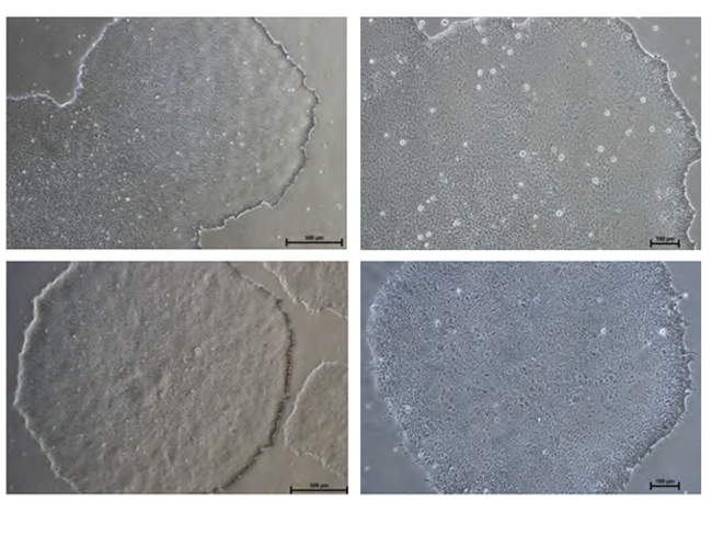
Figure: Normal Colony Morphology. H1 hES cells (top panel) and ACS-1014 hiPS cells (bottom panel) cultured in NutriStem® hPSC XF Medium on Matrigel-coated plates display colony morphologies typical of normal feeder-free hES and hiPS cell cultures, including a uniform colony of tightly compacted cells and distinct colony
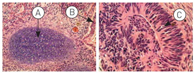
Figure: H9.2 hES cells were cultured for 11 passages in NutriStem® hPSC XF Medium using a human foreskin fibroblast (HFF) feeder layer. The hES cells were subsequently injected into the hind leg muscle of SCID-beige mice for in vitro evaluation of pluripotency. The following tissues from all three germ layers were identified in H&E-stained histological sections of the teratoma 12 weeks post-injection: (A) cartilage (mesoderm), (B) epithelium (endoderm), and (C) neural rosette (ectoderm).
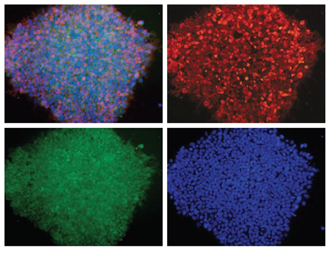
Figure: H1 cell morphology and immunofluorescence analysis of hESC markers red SSEA-4, green OCT4 and blue DAPI. H1 cells stained positive for the expression of pluripotency markers.

Figure: Embryoid bodies (EBs) were generated from H9.2 hES cells cultured for 16 passages in NutriStem® hPSC XF Medium on Matrigel matrix as an evaluation of pluripotency. The pluripotent H9.2 cells were suspended in serum-supplemented medium, where they spontaneously formed EBs containing cells of embryonic germ layers. The following cell types were identified by examination of the histological sections of 14-day-old EBs stained with H&E: (A) neural rosette (ectoderm), (B) neural rosette stained with Tubulin, (C) primitive blood vessels (mesoderm), and (D) megakaryocytes (mesoderm).


